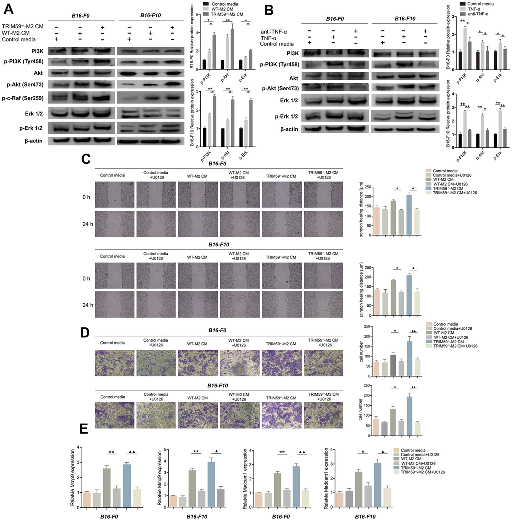
Figure 4.TRIM59-/--M2 macrophage CM activates the PI3K-Akt and ERK pathways in melanoma cells. (A) Western blot detection of signaling proteins related to the PI3K and ERK pathways in B16-F0 and B16-F10 cells treated with control media, WT-M2 CM, or TRIM59-/--M2 CM. Data are represented as mean ± SD. *p<0.05, **p<0.01, compared with the TRIM59-/--M2 CM group. (B) Western blot detection of signaling proteins related to the PI3K and ERK pathways in B16-F0 and B16-F10 cells treated with control media, TNF-α, or a TNF-α antibody. Data are represented as mean ± SD. *p<0.05, **p<0.01, compared with the TNF-α group. (C) Transwell assays results showing the invasive ability of B16-F0 and B16-F10 cells in response to CM from M2 macrophage cultures in the presence of the ERK inhibitor U0126. (D) Wound healing assay results showing the migratory ability of B16-F0 and B16-F10 cells in response to CM from M2 macrophage cultures in the presence of the ERK inhibitor U0126. (E) qRT-PCR evaluation of MMP-9 and Madcam1 expression in B16-F0 and B16-F10 cells exposed to CM from M2 macrophage cultures in the presence of the ERK inhibitor U0126. Data are represented as mean ± SD. *p<0.05, **p<0.01, WT-M2 CM vs. WT-M2 CM plus U0126; ▲p<0.05, ▲▲p<0.01, TRIM59-/--M2 CM vs. TRIM59-/--M2 CM plus U0126.