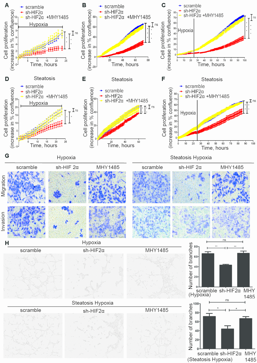
Figure 4.Steatotic HCC growth, migration, invasion and angiogenesis in a hypoxic microenvironment needs HIF-2α. (A–C) Summary graph showing the HCC cell 2D growth rate in hypoxic conditions (A), after 24 hypoxia treatments (B) and all conditions (C). (D–F) Summary graph showing the steatotic HCC cell 2D growth rate under hypoxic conditions (D) after 24 hypoxia treatments (E) and all conditions (F, G). Transwell assays of HCC, HIF-2α-KD HCC, steatotic HCC and HIF-2α-KD steatotic HCC cells under hypoxic conditions in the presence or absence of MHY1485 treatment. h. Representative images of HUVEC tube formation; HUVECs were treated with the supernatant of HCC, HIF-2α-KD HCC, steatotic HCC and HIF-2α-KD steatotic HCC cell cultures.