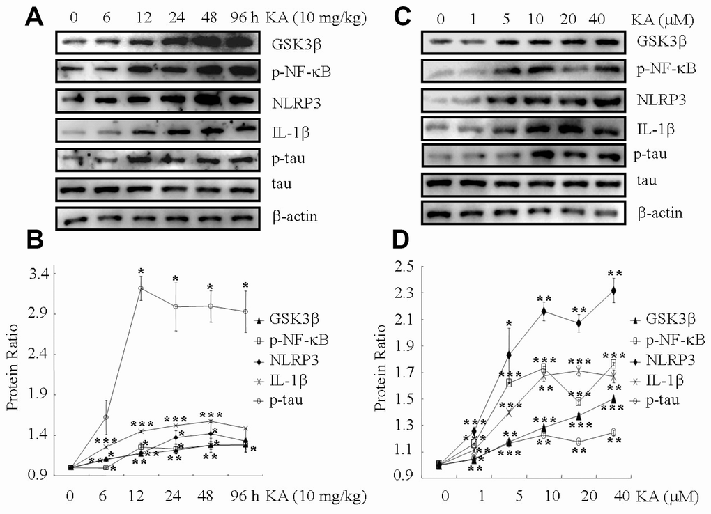
Figure 1.KA augments inflammasome activity and tau phosphorylation in vivo and in vitro. (A, B) Truncation of GSK3β, phosphorylation levels of NF-κB, and the expression levels of NLRP3 and IL-1β as well as the phosphorylation of tau in the KA-treated mouse brain at different time points. (C, D) Truncation of GSK3β, phosphorylation levels of NF-κB, and the expression levels of NLRP3 and IL-1β as well as the phosphorylation of tau in KA-treated mixed cells. The optical density of bands in western blots was analyzed by Image J software (*P < 0.05, **P < 0.01, ***P < 0.001 vs. controls; the significant differences from the respective values were determined by one-way analysis of variance test. N = 3 for western blotting).