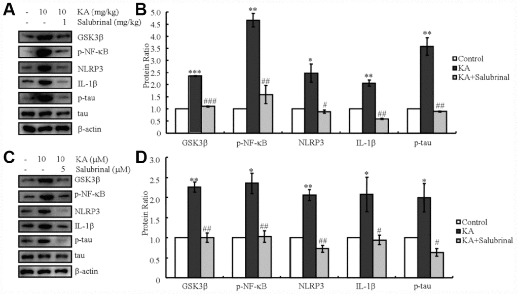
Figure 4.KA-induced tau hyperphosphorylation is highly correlated with ER stress-activated inflammasome. (A, B) Truncation of GSK3β, phosphorylation levels of NF-κB, and the expression levels of NLRP3 and IL-1β as well as the phosphorylation of tau were determined by western blots of samples from MAPT Tg mice treated with KA (10 mg/kg) and/or salubrinal (1 mg/kg) together with KA (10 mg/kg). The KA group was given i.p. injection of 10 mg/kg KA. The salubrinal+KA group mice were additionally given i.p. injections of 1 mg/kg Bay11-7082. Both groups were assessed after 48 h. (C, D) Truncation of GSK3β, phosphorylation levels of NF-κB, and the expression levels of NLRP3 and IL-1β as well as the phosphorylation of tau were determined by western blots in cells treated with KA (10 μM) and/or salubrinal (5 μM) together with KA (10 μM). The KA group was treated with 10 μM KA. The salubrinal+KA group was additionally treated with 5 μM salubrinal. Both groups were assessed after 48 h. The optical density of the bands in western blots was analyzed by Image J software (*P < 0.05; **P < 0.01; ***P < 0.001 vs. the control group; #P < 0.05; ##P < 0.01; ###P < 0.001 vs. the KA-only group).