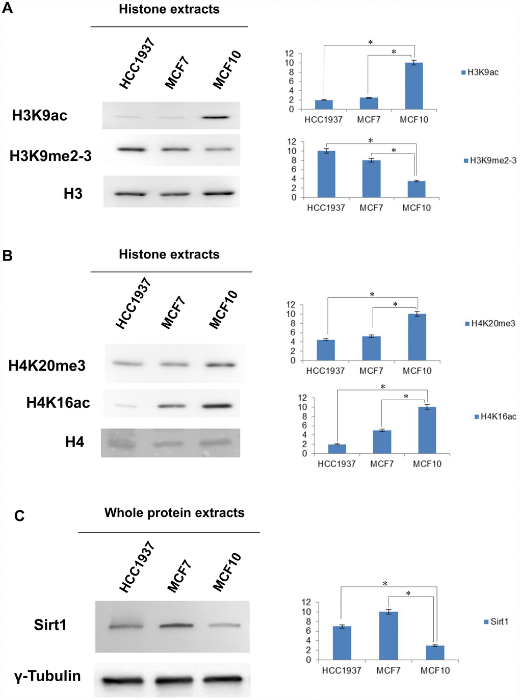
Figure 3.Western blot and densitometry analysis relative to the expression of histone marks and SIRT1 in MCF10, MCF7 and HCC1937 cells. (A) Western blot and densitometry analysis relative to the expression of histone marks in MCF10, MCF7 and HCC1937 cells. (A) H3K9ac, H3K9me2-3, PMTs signal were normalized against the total level of histone H3. (B) H4K20me3, H4K16Ac, PMTs signal were normalized against the total level histone H4. (C) Western blot and densitometry analysis of the expression levels of SIRT1 in whole protein extracts from MCF10, MCF7 and HCC1937 cells. A goat polyclonal anti-γ-Tubulin antibody was used (C-20) to confirm an equal loading of proteins. The assays were repeated in three independent biological replicates and statistically significant differences were determined using one-way ANOVA followed by Dunnett's multiple comparisons test. Data are expressed as mean ± SEM (N =3), p-value <0.05.