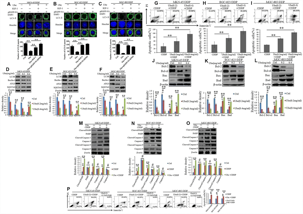
Figure 7.Ubenimex inhibits autophagy and activates CDDP-induced apoptosis in GC cells via suppressing activation of the CD13/EMP3/PI3K/AKT/NF-κB pathway. (A–C) CDDP-resistant GC cells were pre-transfected with pEGFP-N1-EMP3 for 24 h or pre-stimulated with IGF-1 (10 ng/mL) for 8 h, and then treated with Ubenimex (0.2 mg/mL) for another 24 h. LC3-II distribution in MKN-45/DDP (A), BGC-823/DDP (B) and MGC-803/DDP (C) cells were determined via a confocal microscopy, and were represented as the stained granules (upper panels). The number of LC3-II puncta with the means±SD (bottom panels) were also calculated (bottom panels). **P<0.01. (D–F) CDDP-resistant GC cells were treated with Ubenimex (0.2 or 0.4 mg/mL) for 24 h, expression of autophagy-related markers in MKN-45/DDP (D), BGC-823/DDP (E) and MGC-803/DDP (F) cells were identified via Western blot assay. (G–I) CDDP-resistant GC cells were pre-stimulated with Ubenimex, and then treated with CDDP. Apoptosis in MKN-45/DDP (G), BGC-823/DDP (H) and MGC-803/DDP cells (I) were evaluated using Annexin V/PI staining. (J–L) Indicated cells were treated with Ubenimex (0.2 or 0.4 mg/mL) for 24 h, and then stimulated with CDDP (20 μmol/L) for another 48h, the expression of apoptosis related proteins in MKN-45/DDP cells (J), BGC-823/DDP (K) and MGC-803/DDP cells (L) were detected using Western blot assay. (M–O) CDDP-resistant GC cells were pre-treated with Ubenimex (0.2 mg/mL) for 24 h, followed by the stimulation with CDDP (20 μmol/L) for another 48 h. The expression of total and cleaved PARP, Caspase-3 and Caspase-9 in MKN-45/DDP cells (M), BGC-823/DDP (N), and MGC-803/DDP cells (O) were examined by Western blot assay. (P) CDDP-resistant GC cells were pre-stimulated with Ubenimex, and they were treated with Z-VAD-FMK (50μM) for another 2 h before CDDP administration. Cell apoptosis were evaluated using Annexin V/PI staining. For cell apoptosis analysis, data are demonstrated as the representatives (upper or left panels), as well as the proportions of apoptotic cells with the means±SD (bottom or right panels) from three independent experiments, *P<0.05 and **P<0.01. For Western blot assay, data are displayed as the representatives (upper panels) and the means±SD (bottom panels). *P<0.05, **P<0.01and #P>0.05.