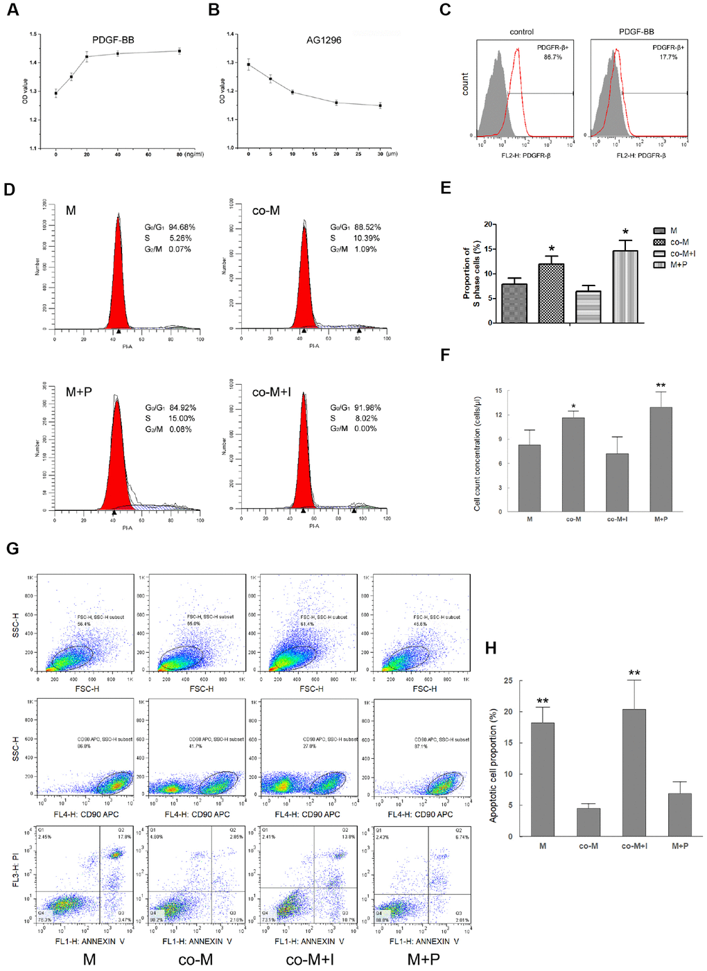
Figure 2.Effect of PDGF-BB/PDGFR-β signaling on viability of MSCs. MSCs were treated with PDGF-BB at concentrations of 10, 20, 40 or 80ng/ml. CCK-8 assay revealed maximal proliferation of MSCs induced with PDGF-BB at concentration of 20 ng/ml or more. Results are mean ± SD from three independent experiments (A). co-MSCs were treated with AG1296 (PDGFR-β inhibitor), and CCK-8 assay revealed maximal inhibition of proliferation at concentration of 20μm or more. Results are mean ± SD from three independent experiments (B). Flow cytometry revealed that after treated with 20ng/ml PDGF-BB, detection of PDGFR-β+ MSCs decreased due to combination of PDGF-BB and PDGFR-β (C). The cell cycle analysis noted that compared with M and co-M+I groups, the proportion of cells in S phase significantly increased in either co-M or M+P group. Results are mean ± SD from three independent experiments (D, E). Cell count concentration was assessed using the CountBright with flow cytometry. The result was consistent with the cell cycle. Results are mean ± SD from three independent experiments (F). Cell apoptosis detected with flow cytometry revealed that compared with M and co-M+I groups, the proportion of apoptotic cells significantly decreased in either co-M or M+P group (G, H). *P<0.05 vs. M or co-M+I group, **P<0.01 vs. M or co-M+I group.