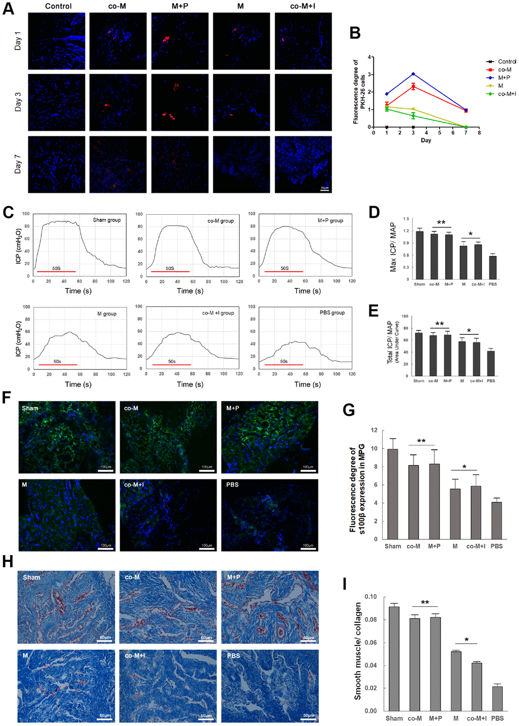
Figure 4.In vivo viability and nerve regenerative capacity of MSCs induced with PDGF-BB/PDGFR-β signaling. PKH26-labled cells (Red) were detected in the MPG and cavernous nerve 1, 3, 7 and 14 days after implantation. PKH26-labled cells were detectable within 3 days after implantation in either M or co-M+I group. However, they were durable and detectable until 7 days after implantation in both co-M and M+P groups. They were undetectable 14 days after implantation in all groups (data not shown). Image J revealed a stronger fluorescence degree of PKH-26 cells in the co-M and M+P groups than either M or co-M+I group (B). Rats per group: n=2. Scale bar=50μm. Transplantation of different groups of MSCs restored erectile function in CNI rats (C–E). Representative ICP responses for the sham, M, co-M, co-M+I, M+P and PBS group 2 weeks after treatment of stem cells (C). Compared with the PBS group, treatment of MSCs significantly increased the mICP/MAP and tICP/MAP ratios. Moreover, the data of the co-M and M+P group were better than the M or co-M+I group (D, E). Each bar represents mean ± SD (n=5 animals per group). *P<0.05 vs. PBS group; **P<0.01 vs. PBS group and P<0.05 vs. M and co-M+I group. MAP=mean arterial pressure. The expressions of neural marker S100β in MPG were detected 2 weeks after treatment of stem cells (F). The ratio of smooth muscle to collagen in penis was assessed with Masson’s trichrome staining (H). Quantitative analysis was performed using Image J (G, I). Each bar represented means ± SD (n=5 animals per group). *P<0.05 vs. PBS group; **P<0.01 vs. PBS group and P<0.05 vs. M and co-M+I group.