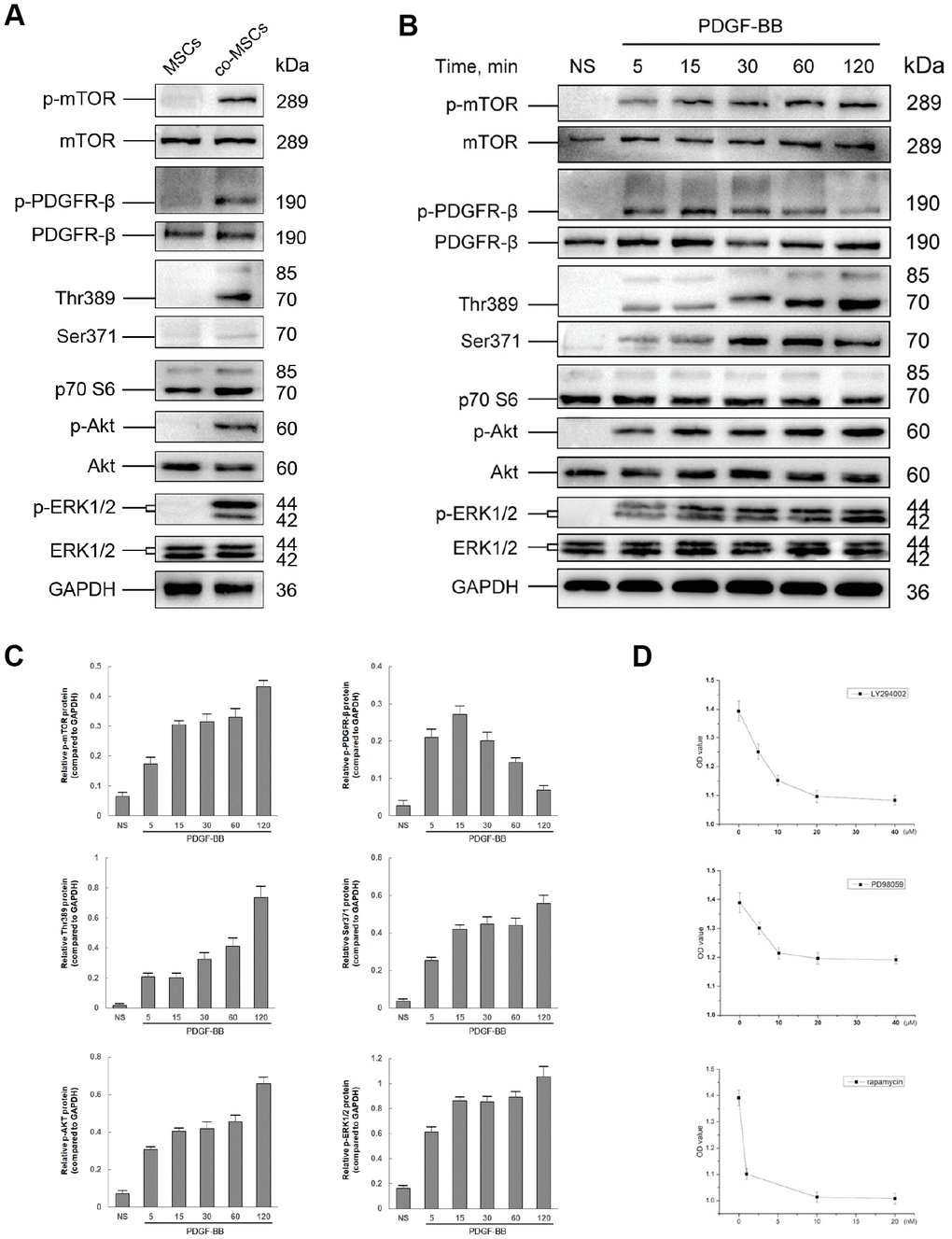
Figure 5.The role of PI3K/Akt and MEK/Erk pathway in PDGF-BB-induced viability of MSCs. Either co-cultured with EPCs or 20 ng/ml PDGF-BB was applied to induced viability of MSCs and the status in phosphorylation of both PI3K/Akt and MEK/Erk pathways was assessed by western blot. Co-cultured with EPCs, the co-MSCs revealed phosphorylation of PDGFR-β, Akt, mTOR, p70 S6 Kinase (Thr389 and Ser371) and ERK ½ (A). Similarly, after induced with PDGF-BB, PDGFR-β, Akt, mTOR, p70 S6 Kinase and ERK ½ was rapidly phosphorylated (B). The densitometry of western blots was evaluated by Image J and the results revealed that phosphorylation of PDGFR-β reached peak at 15 min and gradually returned to basal level. However, the phosphorylation of downstream pathway factors consistently existed. Results are mean ± SD from three independent experiments (C). Different concentrations of LY294002 (PI3K inhibitor), PD98059 (Erk inhibitor) or rapamycin (mTOR inhibitor) were pre-treated to MSCs 1h before treated with PDGF-BB. CCK-8 cell proliferation test revealed that the optimal concentration of LY294002, PD98059 and rapamycin was 20μM, 10μM and 10nM, respectively. Results are mean ± SD from three independent experiments (D).