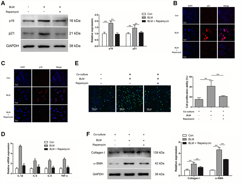
Figure 4.Rapamycin could suppress epithelial cell senescence and fibroblast activation via impairing the production of SASP. (A–E) MLE-12 cells were treated with bleomycin (BLM), followed by treatment with or without rapamycin for 3 days. (A) The expression of p16 and p21 were measured by Western blot. The expression levels were quantified with ImageJ (n = 3). GAPDH was used as a loading control, **P < 0.01. (B, C) The protein levels of p16 and p21 were detected by immunofluorescence. (D) The mRNA levels of IL-1β, IL-6, IL-8 and TNF-α were determined by Q-PCR, ** P < 0.01 vs. Con and ## P < 0.01 vs. BLM. (E, F) MLE-12 cells were treated as in Figure 4A and co-cultured with pulmonary fibroblasts in fresh medium for another 3 days. (E) The proliferation ability of pulmonary fibroblasts were measured by EdU assay. The percentage of proliferating cells was calculated by ImageJ, **P < 0.01. (F) The expression of α-SMA and collagen I were detected by Western blot. The expression levels were quantified with ImageJ (n = 3). GAPDH was used as a loading control, **P < 0.01.