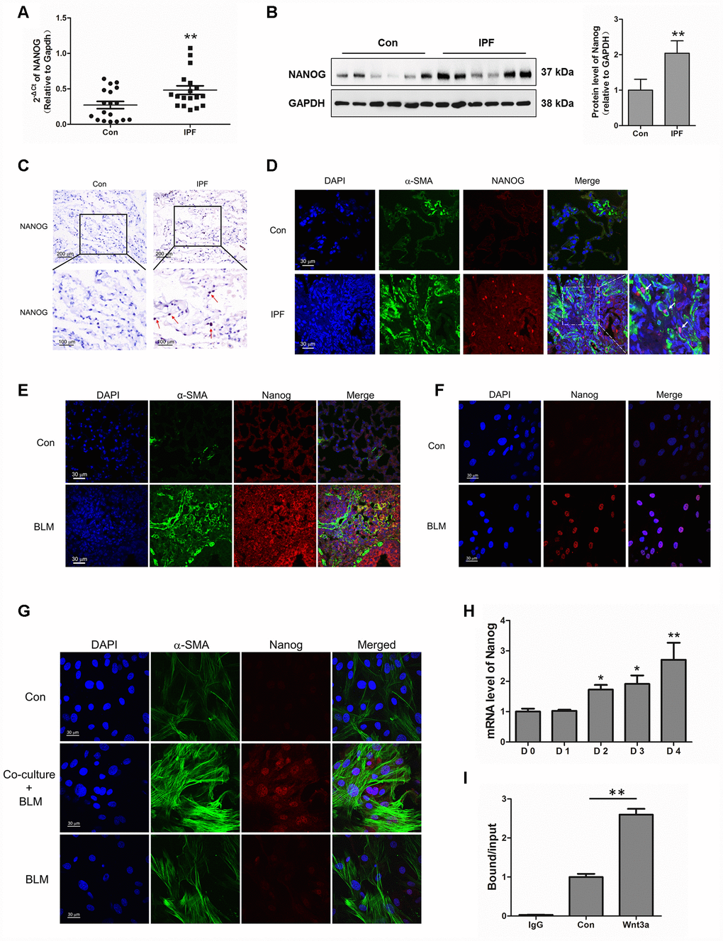
Figure 5.Aberrantly expressed Nanog in activated pulmonary fibroblasts and fibrotic lung tissues were mediated by Wnt/β-catenin. (A–C) The expression of Nanog in lung tissues derived from patients with idiopathic pulmonary fibrosis (IPF) was determined by Q-PCR (A), Western blot (B) and immunohistochemical analysis (C), ** P < 0.01 vs. Con. (D) The lung tissues of patients with IPF were double stained with α-SMA and Nanog by immunofluorescence. (E) The lung tissues derived from pulmonary fibrosis mouse models were double stained with α-SMA and Nanog via immunofluorescence. (F) The expression of Nanog in pulmonary fibroblasts isolated from fibrotic mouse lung tissues were measured by immunofluorescence. (G) Cells were treated as in Figure 3D. Pulmonary fibroblasts were double stained with α-SMA and Nanog by immunofluorescence. (H, I) Pulmonary fibroblasts were treated with Wnt3a for various durations. (H) The mRNA level of Nanog was detected by Q-PCR, * P < 0.05 and ** P < 0.01 vs. D0. (I) ChIP assays were performed by using chromatin isolated from Wnt3a treated pulmonary fibroblasts. The final DNA extracts were analysed by Q-PCR, ** P < 0.01.