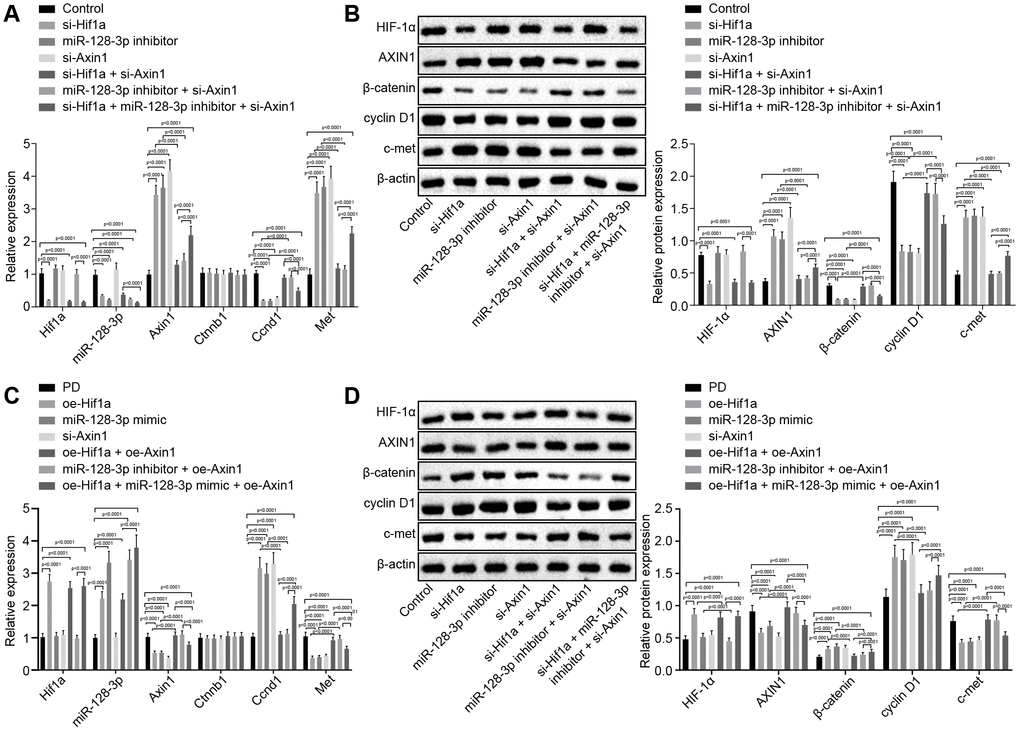
Figure 3.The expression pattern of HIF-1α/miR-128-3p/AXIN1 and the Wnt/β-catenin signaling pathway-related proteins (β-catenin, cyclin D1 and c-met) in hippocampal neurons of normal mice (n = 10) and in the MPTP-lesioned mouse model of PD (n = 30) after transduction. (A) The expression levels of Hif1a, miR-128-3p, Axin1, Ctnnb1, Ccnd1, and Met in primary hippocampal neurons of normal mice after treated with si-Hif1a, miR-128-3p inhibitor, or si-Axin1, determined by RT-qPCR. (B) The protein band patterns and the protein levels of HIF-1α, AXIN1, β-catenin, cyclin D1, and c-met normalized to β-actin in primary hippocampal neurons of normal mice after treatment with si-Hif1a, miR-128-3p inhibitor, or si-Axin1, all as determined by western blot analysis. * p < 0.05 vs. the control group (primary hippocampal neurons of normal mice); # p < 0.05 vs. the si-Hif1a group (hippocampal neurons of normal mice treated with si-Hif1a); & p < 0.05 vs. the miR-128-3p inhibitor group (hippocampal neurons of normal mice treated with miR-128-3p inhibitor); @ p < 0.05 vs. the si-Hif1a + si-Axin1 group (hippocampal neurons of normal mice treated with si-Hif1a + si-Axin1); $ p < 0.05 vs. the miR-128-3p inhibitor + si-Axin1 group (hippocampal neurons of normal mice treated with miR-128-3p inhibitor + si-Axin1). (C) The expression levels of Hif1a, miR-128-3p, Axin1, Ctnnb1, Ccnd1, and Met in primary hippocampal neurons cultured from the MPTP-lesioned mouse model of PD after treatment with oe-Hif1a, miR-128-3p mimic, or oe-Axin1, as determined by RT-qPCR. (D) The protein band patterns and the protein levels of HIF-1α, AXIN1, β-catenin, cyclin D1, and c-met normalized to β-actin in primary hippocampal neurons of the MPTP-lesioned mouse model of PD after treatment with oe-Hif1a, miR-128-3p mimic, or oe-Axin1, as determined by western blot analysis. * p < 0.05 vs. the PD group (primary hippocampal neurons of MPTP-lesioned mouse model of PD); # p < 0.05 vs. the oe-Hif1a group (hippocampal neurons of MPTP-lesioned mouse model of PD treated with oe-Hif1a); & p < 0.05 vs. the miR-128-3p mimic group (hippocampal neurons of MPTP-lesioned mouse model of PD treated with miR-128-3p mimic); @ p < 0.05 vs. the oe-Hif1a + oe-Axin1 group (hippocampal neurons of MPTP-lesioned mouse model of PD treated with oe-Hif1a + oe-Axin1); $ p < 0.05 vs. the miR-128-3p mimic + oe-Axin1 group (hippocampal neurons of MPTP-lesioned mouse model of PD treated with miR-128-3p mimic + oe-Axin1). The experiment was repeated tree times independently.