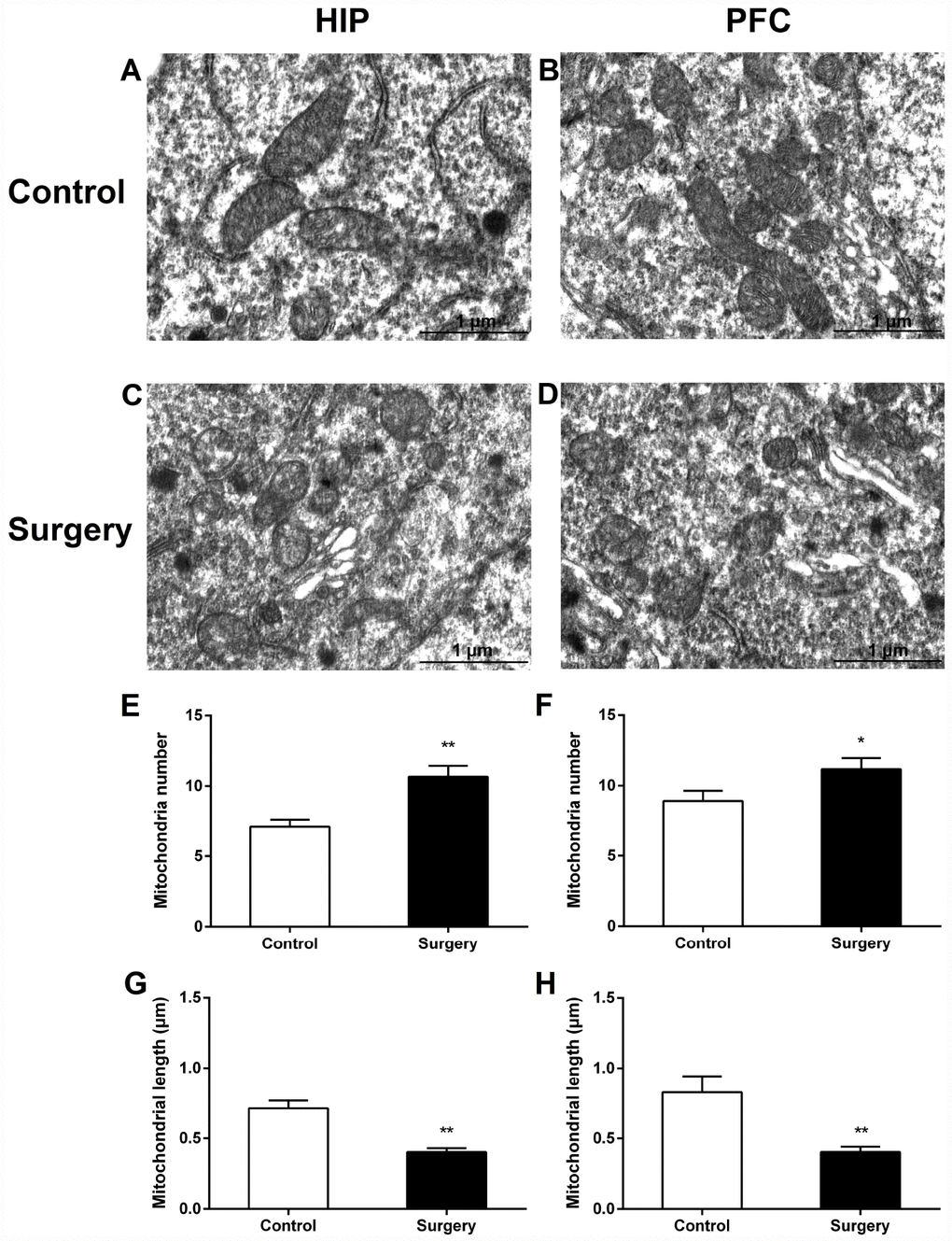
Figure 7.Surgery/Anesthesia caused ultrastructural changes in the mitochondria of hippocampal and prefrontal cortex neurons in aged mice at 24 hours postoperatively. Mitochondria in the cytoplasm of hippocampal (A) and prefrontal cortex (B) neurons from the control mice resemble long tubules with intact outer and inner membranes and numerous cristae tightly packed in healthy looking matrix. The number of mitochondria in the cytoplasm of hippocampal (C and E) and prefrontal cortex (D and F) neurons from mice in the Surgery/Anesthesia group were increased significantly. Compared to the control condition, Surgery/ Anesthesia decreased the mitochondrial length in the hippocampus (G) and prefrontal cortex (H) at 24 hours postoperatively. The mitochondria in the Surgery/Anesthesia group were small, round, and displayed globular morphology. Although the outer and inner membranes appeared somewhat intact, the cristae seemed distorted and difficult to discern. Scale bar: 1 μm. The data are plotted as the mean ± standard error of the mean for each group (n = 3). *p < 0.05 and **p < 0.01, compared to control.