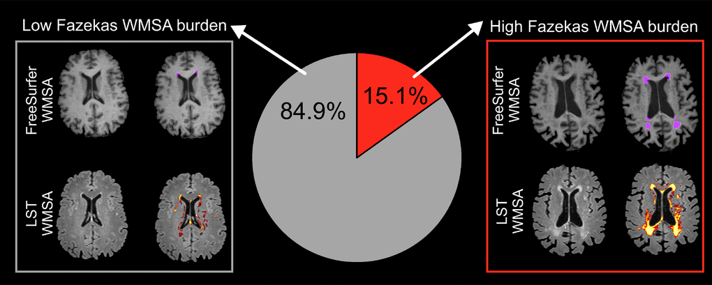
Figure 1.Prevalence of low and high Fazekas WMSA burden. Low WMSA burden was defined as Fazekas scores 0 (i.e. absence of WMSA) or 1 (i.e. punctate WMSA). High WMSA burden was defined as Fazekas scores 2 (i.e. early confluent WMSA) and 3 (i.e. WMSA in large confluent areas). The gray box illustrates automatic segmentations of WMSA by FreeSurfer (first row) and LST (second row), for a representative subject with low Fazekas WMSA burden. The red box illustrates automatic segmentations of WMSA by FreeSurfer (first row) and LST (second row), for a representative subject with high Fazekas WMSA burden. WMSA: White matter signal abnormalities; LST: Lesion segmentation tool.