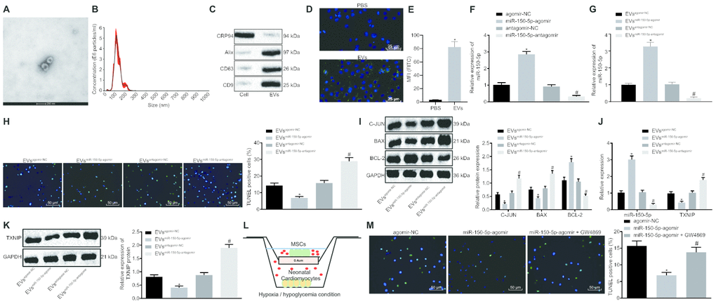
Figure 4.Cardiomyocyte apoptosis is suppressed by MSCs-derived EVs carrying miR-150-5p. (A) The images captured under transmission electron microscopy. (B) EV size measured by nanoparticle tracking analysis. (C) The expression of EV markers (CD9, CD63, Alix and GRP94) detected by Western blot analysis. (D) EV internalization detected by immunohistochemistry (400 ×). (E) PKH67-staining (FITC wavelength) in neonatal cardiomyocytes detected by flow cytometric analysis. (F) The expression of miR-150-5p in MSCs determined by RT-qPCR. (G) The expression of miR-150-5p in EVs determined by RT-qPCR. (H) neonatal cardiomyocyte apoptosis detected by TUNEL staining (200 ×). (I) The protein expression of apoptosis-related factors (c-Jun, Bax and Bcl-2) normalized to GAPDH determined by Western blot analysis. (J) The expression of miR-150-5p and TXNIP in neonatal cardiomyocytes determined by RT-qPCR. (K) TXNIP expression in neonatal cardiomyocytes normalized to GAPDH determined by Western blot analysis. (L) Transwell co-culture system under hypoxia/hypoglycemia condition. (M) Neonatal cardiomyocyte apoptosis detected by TUNEL staining (200 ×); * p < 0.05 vs. the agomir-NC group (cells treated with agomir-NC); # p < 0.05 vs. the miR-150-5p-agomir group (cells treated with miR-150-5p-agomir). * p < 0.05 vs. the agomir-NC group (cells treated with agomir-NC) or the EVagomir-NC group (cells treated with EVagomir-NC); # p < 0.05 vs. the antagomir-NC group (cells treated with antagomir-NC) or the EVantagomir-NC group (cells treated with EVantagomir-NC). Measurement data were presented as mean ± standard deviation. Comparison among multiple groups was analyzed by one-way analysis of variance, followed by Tukey’s post hoc test. The cell experiment was repeated 3 times independently.