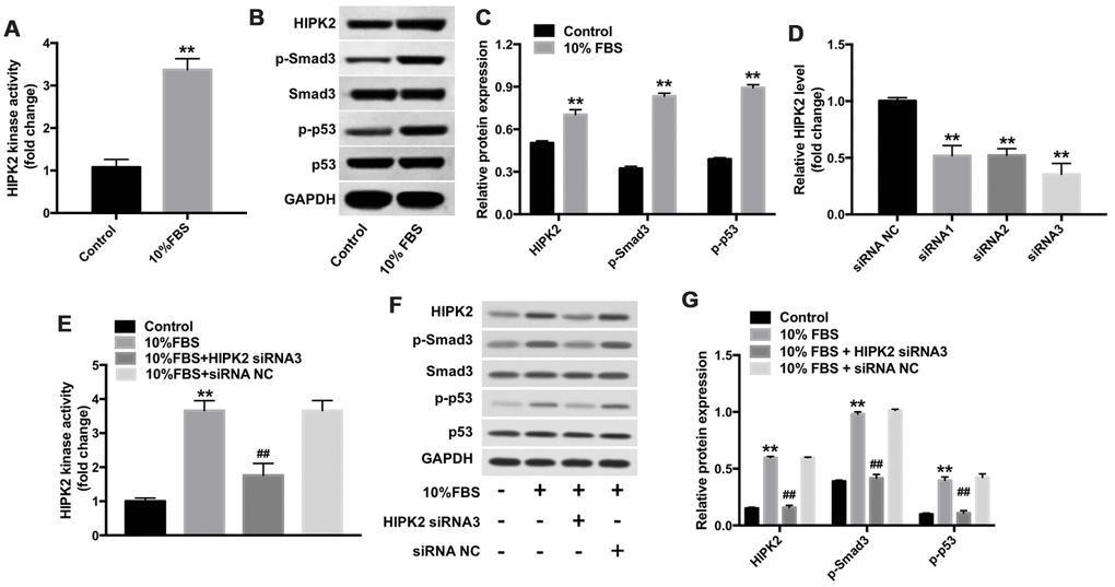
Figure 4.Downregulation of HIPK2 suppresses FBS-induced differentiation of rabbit corneal keratocytes into myofibroblasts. (A) Histogram plot shows HIPK2 kinase activity in rabbit corneal keratocytes grown in DMEM/F12 medium with and without 10% FBS. (B, C) Western blot analysis shows levels of HIPK2, Smad3, p-Smad3, p53, and p-p53 in rabbit corneal keratocytes grown in DMEM/F12 medium with and without 10% FBS. GAPDH was used as internal control. Histogram plot shows levels of HIPK2, p-Smad3, and p-p53 relative to GAPDH, Smad3, and p53, respectively. (D) QRT-PCR analysis shows relative HIPK2 mRNA levels in rabbit corneal keratocytes transfected with NC-siRNA, HIPK2-siRNA1, HIPK2-siRNA2, and HIPK2-siRNA3 for 48 h. β-actin mRNA levels were used for normalization. (E) HIPK2 kinase activity in rabbit corneal keratocytes, transfected with NC-siRNA or HIPK2-siRNA3, and grown in DMEM/F12 medium with 10% FBS for 48 h. (F, G) Western blot analysis shows HIPK2, Smad3, p-Smad3, p53, and p-p53 levels in rabbit corneal keratocytes, transfected with NC-siRNA or HIPK2-siRNA3, and grown in DMEM/F12 medium with 10% FBS for 48 h. GAPDH was used as internal control. Histogram plot shows levels of HIPK2, p-Smad3, and p-p53 relative to GAPDH, Smad3, and p53, respectively. ** denotes P < 0.01 as compared with the control group; ## denotes P < 0.01 as compared with the 10% FBS + NC-siRNA group.