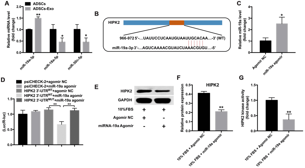
Figure 5.Mir-19a directly binds to the HIPK2-3’UTR. (A) QRT-PCR analysis shows miR-19a, miR-18a-5p and miR-30c-5p levels in ADSCs and ADSCs-Exo. ** denotes P < 0.01 in comparison with the ADSC group. (B) Targetscan analysis shows the sequence, 5’-UGCACA-3’, as the predicted target site of miR-19a in the 3’UTR region of the HIPK2 gene between 966-972 nucleotides. (C) QRT-PCR analysis shows levels of miR-19a in the rabbit corneal keratocytes transfected with miR-19a-agomir or NC-agomir for 48 h. ** denotes P < 0.01 in comparison with the agomir NC group. (D) The dual luciferase reporter assay shows relative luciferase activity in the 293T cells co-tranfected with the plasmids containing HIPK2-WT-3’UTR or HIPK2-MUT-3’UTR and miR-19a agomir. (E, F) Western blotting analysis shows HIPK2 protein expression in rabbit keratocytes, grown in DMEM/F12 medium with 10% FBS, and transfected with miR-19a-agomir or NC-agomir for 48 h. Histogram plot shows HIPK2 protein levels relative to GAPDH. ** denotes P < 0.01 when compared with the 10% FBS + NC-agomir group. (G) Histogram plot shows HIPK2 kinase activity in rabbit keratocytes grown in DMEM/F12 medium with 10% FBS and transfected with miR-19a agomir or NC-agomir for 48 h. ** denotes P < 0.01 as compared with the 10% FBS + NC-agomir group.