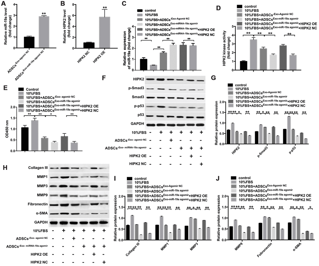
Figure 6.ADSCs-Exo-miR-19a suppresses FBS-induced differentiation of rabbit corneal keratocytes into myofibroblasts by inhibiting HIPK2 expression. (A) QRT-PCR analysis shows miR-19a levels in ADSC-Exo obtained from ADSCs that were transfected with miR-19a-agomir (ADSCs-Exo-miR-19a-agomir) or NC-agomir (ADSCs-Exo-NC-agomir) for 48 h. ** denotes P < 0.01 as compared with the ADSCs-Exo-NC-agomir group. (B) QRT-PCR analysis shows HIPK2 mRNA levels in rabbit corneal keratocytes, transfected with lenti-NC or lenti-HIPK2 for 48 h. β-actin was used as internal control. ** denotes P < 0.01 as compared with the HIPK2-NC group. (C) QRT-PCR analysis shows miR-19a levels in lenti-HIPK2 transfected rabbit corneal keratocytes, grown in DMEM/F12 medium with 10% FBS, and incubated with 100 μg/mL ADSCs-Exo-miR19a-agomir or 100 μg/mL ADSCs-Exo-miR19a-agomir. Exosomes were obtained by ultracentrifugation of the cell culture supernatant of ADSCs that were transfected with miR-19a-agomir or NC-agomir for 48 h. ** denotes P < 0.01. (D) Histogram plot shows HIPK2 kinase activity in lenti-HIPK2 transfected rabbit corneal keratocytes, grown in DMEM/F12 medium with 10% FBS, and incubated with 100 μg/mL ADSCs-Exo-miR19a-agomir or 100 μg/mL ADSCs-Exo-NC-agomir. ** denotes P < 0.01. (E) CCK-8 assay results show viability of lenti-HIPK2 transfected rabbit corneal keratocytes, grown in DMEM/F12 medium with 10% FBS, and incubated with 100 μg/mL ADSCs-Exo-miR19a-agomir or 100 μg/mL ADSCs-Exo-NC-agomir at 0, 2, 48, and 72 h. (F, G) Western blot analysis shows HIPK2, Smad3, p-Smad3, p53, and p-p53 in lenti-HIPK2 transfected rabbit corneal keratocytes, grown in DMEM/F12 medium with 10% FBS, and incubated with 100 μg/mL ADSCs-Exo-miR19a-agomir or 100 μg/mL ADSCs-Exo-NC-agomir. The levels of HIPK2, p-Smad3, and p-p53 proteins are expressed relative to GAPDH, Smad3, and p53, respectively. (H) Western blot analysis of the levels of collagen III, MMP1, MMP3, MMP9, fibronectin and α-SMA proteins in lenti-HIPK2 transfected rabbit corneal keratocytes, grown in DMEM/F12 medium with 10% FBS, and incubated with 100 μg/mL ADSCs-Exo-miR19a-agomir or 100 μg/mL ADSCs-Exo-NC-agomir. GAPDH was used as an internal control. (I, J) Histogram plots show the levels of (I) collagen III, MMP1, and MMP3 and (J) MMP9, fibronectin and α-SMA relative to GAPDH in lenti-HIPK2 transfected rabbit corneal keratocytes, grown in DMEM/F12 medium with 10% FBS, and incubated with 100 μg/mL ADSCs-Exo-miR19a-agomir or 100 μg/mL ADSCs-Exo-NC-agomir. ** denotes P < 0.01.