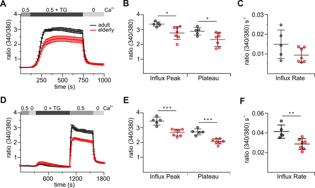
Figure 1.Untouched CD8+ T cells from elderly mice show reduced thapsigargin (TG)-induced Ca2+ signals. (A) Fura2-AM based Ca2+ Imaging with 1 μM TG as stimulus applied in the presence of 0.5 mM [Ca2+]ext of CD8+ T cells (combined Ca2+ protocol) from adult (black, n = 5) and elderly (red, n = 7) mice. The scatter dot plot in (B) displays the corresponding statistics of Ca2+ influx peak and Ca2+ plateau and in (C) the corresponding influx rates. (D) Ca2+ Imaging with 1 μM TG applied in the absence of [Ca2+]ext before re-addition of 0.5 mM Ca2+ (re-addition protocol) of CD8+ T cells from adult (black, n = 5) and elderly (red, n = 7) mice. The scatter dot plot in (E) displays the corresponding statistics of Ca2+ influx peak and Ca2+ plateau and (F) the corresponding influx rates. Ca2+ signalling curves are presented as mean ± SEM. Scatter dot plots are presented as mean ± SD. * p < 0.05, ** p < 0.01, *** p < 0.001, **** p < 0.0001.