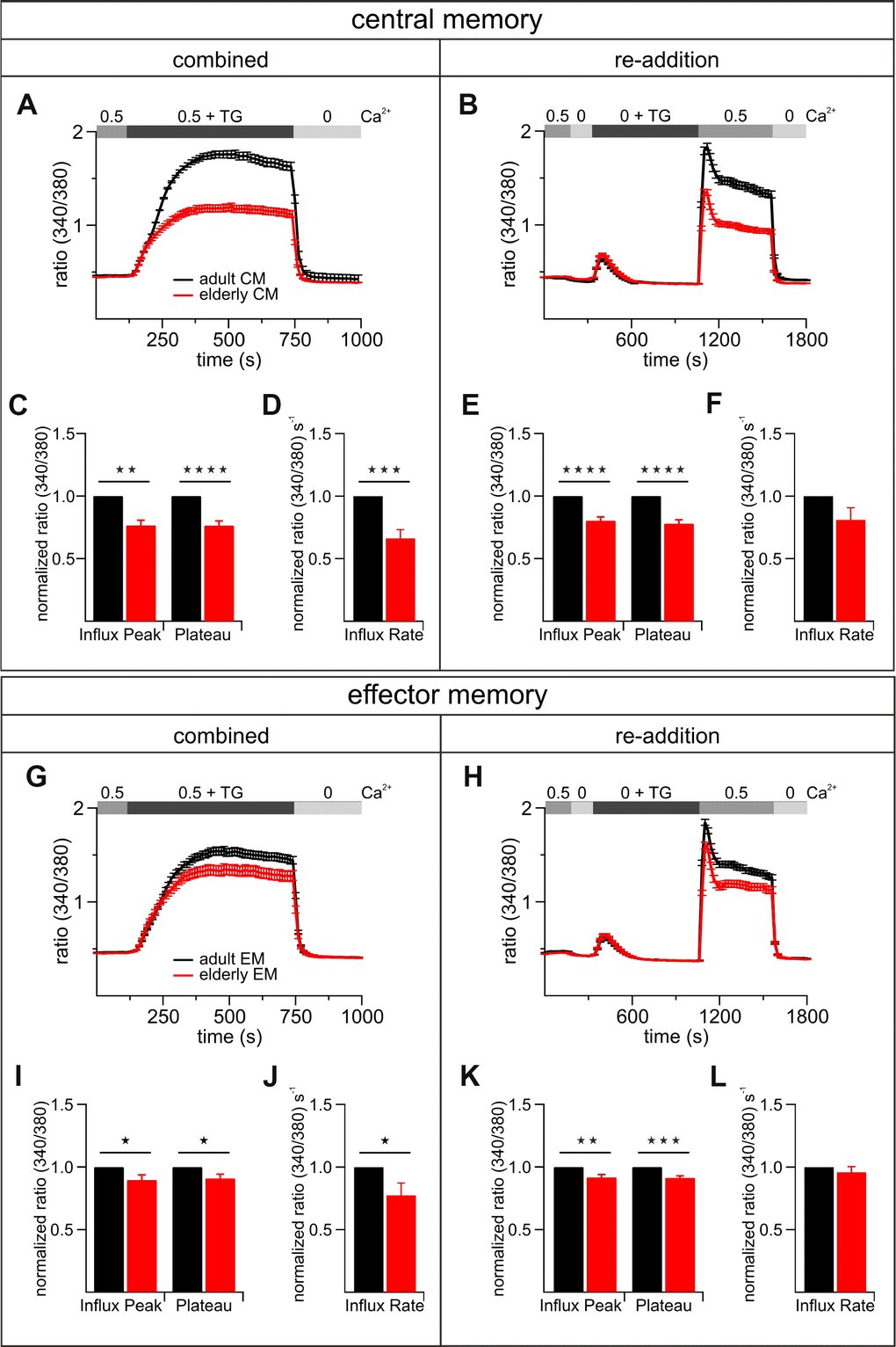
Figure 7.The most abundant subtypes of activated CD8+ T cells exhibit age-related reductions of TG-induced Ca2+ signals. Combined (A) and re-addition (B) protocol of CD8+ central memory T cells (CM) from adult (black, n = 6) and elderly (red, n = 6) mice. The bar graphs in (C) and (E) display the corresponding statistics of Ca2+ influx peak and Ca2+ plateau and in (D) and (F) the corresponding influx rates of combined and re-addition protocol, respectively. Combined (G) and re-addition (H) protocol of CD8+ effector memory T cells (EM) from adult (black, n = 6) and elderly (red, n = 6) mice. The scatter dot plots in (I) and (K) display the corresponding statistics of Ca2+ influx peak and Ca2+ plateau and in (J) and (L) the corresponding influx rates of combined and re-addition protocol, respectively. Ca2+ signalling curves show one exemplary out of six measurements with equal tendencies of central and effector memory cells of adult and elderly mice as mean ± SEM. Bar graphs show values of CD8+ T cells from elderly mice normalized to the values of CD8+ T cells from adult mice as mean ± SEM. * p < 0.05, ** p < 0.01, *** p < 0.001, **** p < 0.0001.