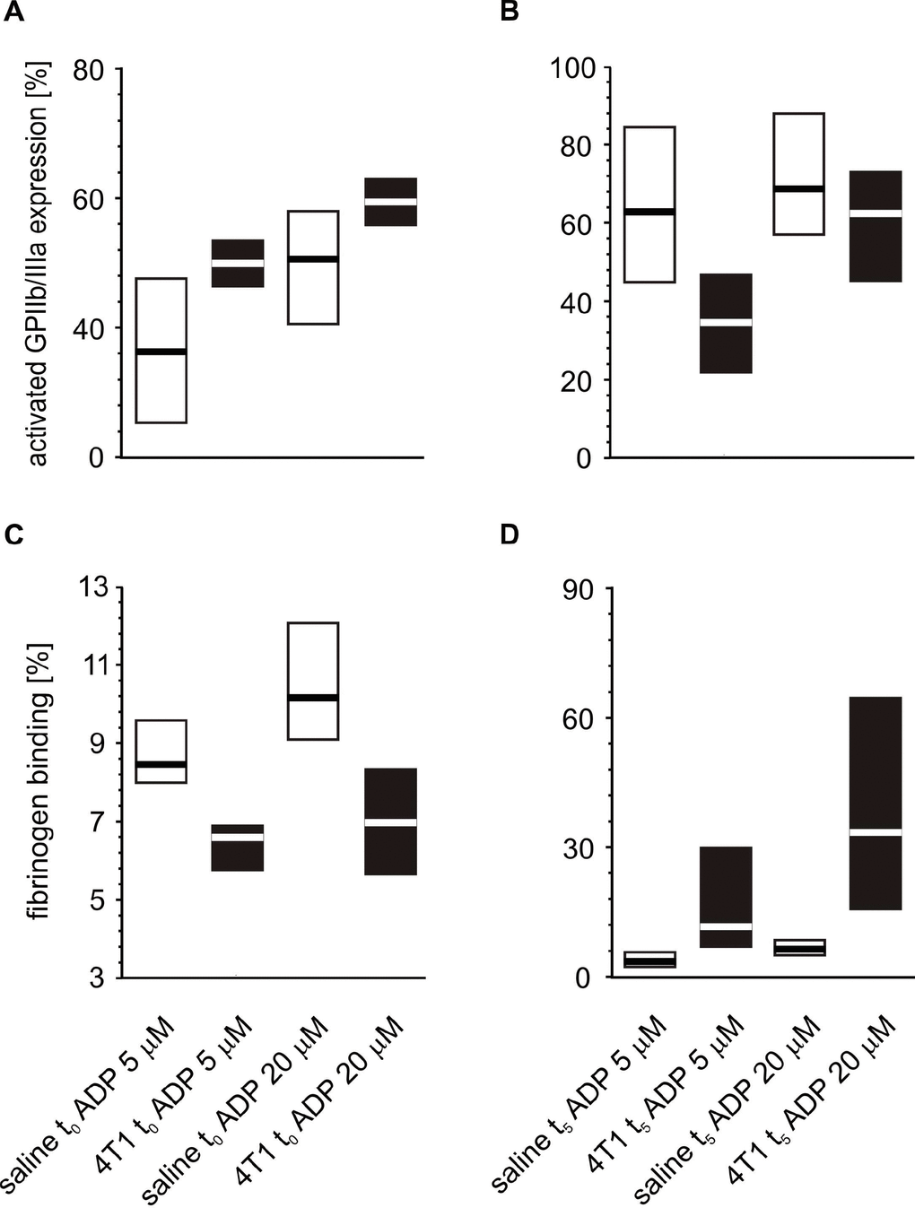
Figure 7.Expressions/bindings of selected platelet surface membrane activation markers on ADP-activated blood platelets in mice injected with 4T1 cancer cells or saline. Results are presented as median (horizontal line) and interquartile range (box) (n = 8). The expressions of the active form of GPIIb/IIIa (A, B) and binding of endogenous fibrinogen (Fg) (C, D) on platelets stimulated with ADP (5 or 20 μM) were measured using flow cytometry in non-fixed ‘washed blood’ withdraw immediately (t0) (A, C) or after 5 weeks (t5) (B, D) from the injection of 4T1-cells or saline. Results are expressed as the percent fraction of platelets positive for a given activation marker. More experimental details are given in the Materials and methods section. The statistical significance of differences, estimated with Kruskal-Wallis test followed by post hoc Conover-Inman all-pairwise comparisons or two-way ANOVA followed by Tukey’s multiple comparisons test, was: active form of GPIIb/IIIaADP5μM, P1,α < 0.01, 4T1 t0 > saline t0; P1,α < 0.01, 4T1 t5 < saline t5; active form of GPIIb/IIIaADP20μM, P1,α < 0.01, 4T1 t0 > saline t0; P1,α < 0.01, 4T1 t5 < saline t5; FgADP5μM, P1,α < 0.01, 4T1 t0 < saline t0; P1,α < 0.01, 4T1 t5 < saline t5; FgADP20μM, P1,α < 0.01, 4T1 t0 < saline t0; P1,α < 0.01, 4T1 t5 < saline t5.