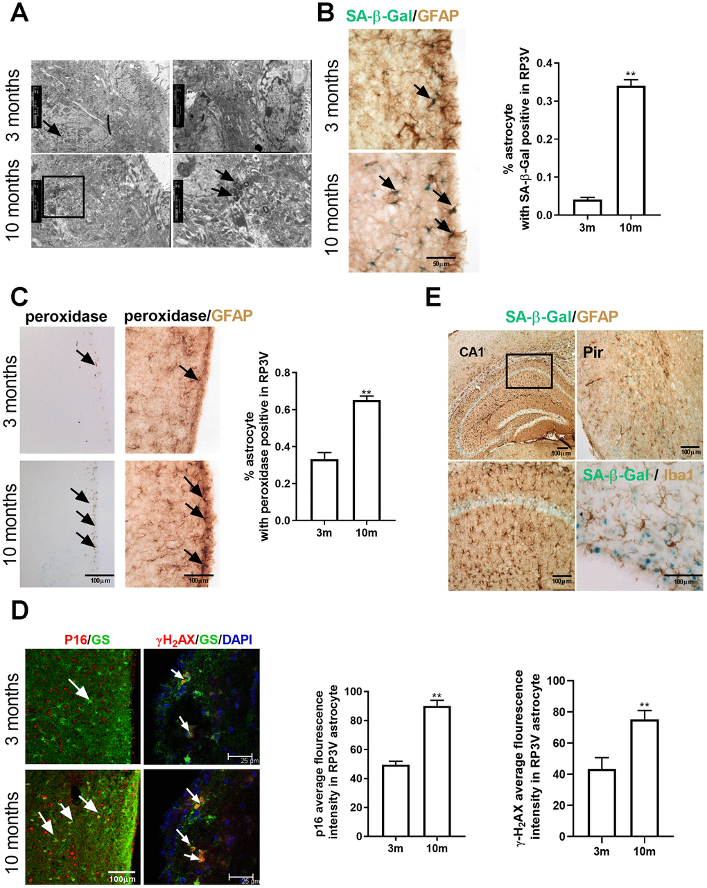
Figure 1.Astrocytes within the hypothalamic RP3V accumulates senescence-related markers with increasing age. (A) The lipofuscin deposition by transmission electron microscopy in hypothalamic astrocytes of female C57BL/6J mice at the age of 3 months and 10 months. Black arrows represent the lipofuscin deposition. (B) Dual-label immunohistochemistry of astrocytes by GFAP staining (brown) and by SA-β-Gal staining (blue) in 3-month-old mice (n=5) and 10-month-old mice (n=5), black arrows representing SA-β-Gal –positive astrocytes, scale bar=50μm. (C) Peroxidase staining (brown) in the astrocytes of RP3V (left), black arrows representing peroxidase. GFAP (black) and peroxidase (brown) double staining in astrocytes of RP3V in the hypothalamus of 3-month-old mice (n=5) and 10-month-old mice (n=5), black arrows representing peroxidase–positive astrocytes, scale bar =100μm. (D) Dual-label immunofluorescence showing astrocytes (green) with p16 (red) in young (n=5) and middle-aged mice (n=5), white arrows representing p16–positive astrocytes, scale bar=100μm (left). Dual-label immunofluorescence showing astrocytes (green) with γ-H2AX (red) in young (n=5) and middle-aged mice (n=5), white arrows representing γH2AX–positive astrocytes, scale bar=25μm (right). (E) Dual-label immunohistochemistry showing astrocytes (brown) with SA-β-Gal staining (blue) in 10-month-old mouse cortex (left picture) and hippocampal (top right picture). Dual-label immunohistochemistry showing microglia (brown) with SA-β-Gal staining (blue) in RP3V of 10-month-old mice (bottom right picture). Scale bar=100μm. The p-value was determined by Student’s t test,** p< 0.01.RP3V, i.e. rostral periventricular area of the third ventricle; GS, i.e. glutamine synthetase.