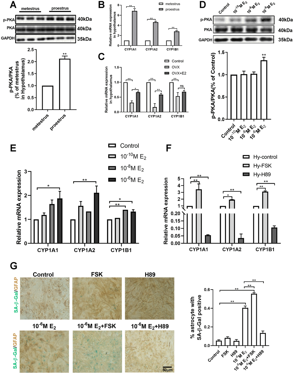
Figure 4.PKA-CYP signaling mediates estradiol-induced senescence of hypothalamic astrocytes. (A) The expression of PKA and p-PKA in hypothalamus during proestrus and metestrus at 3 months of age as determined by Western blotting (n = 6). (B) Effects of estrous cycle on the mRNA expression of CYP1A1, CYP1A2 and CYP1B1 gene in the hypothalamus as determined by qPCR. Metestrus vs. proestrus, n=3 (upper). (C) Effects of estradiol on the mRNA expression of CYP1A1, CYP1A2 and CYP1B1 gene in the hypothalamic tissue as determined by qPCR. OVX group vs. control group, OVX group vs. OVX+E2 group, n=3. The p-value of (A–C) was determined by Student’s t test,*p<0.05,** p< 0.01. (D) Expression of PKA and p-PKA in primary cultured hypothalamic astrocytes with the intervention of different estradiol concentrations as determined by Western blotting (n = 5). (E) Effects of estradiol on the mRNA expression of CYP1A1, CYP1A2 and CYP1B1 gene in primary cultured hypothalamic astrocytes with the intervention of different estradiol concentrations, as compared with the control, n= 3. (F) Effects of PKA activator (Forskolin, 10μM, 24h) and inhibitor (H89, 30μM, 24h) on the mRNA expression of CYP1A1, CYP1A2 and CYP1B1 gene in the primary cultured hypothalamic astrocytes, as compared with the control, n=3. (G) Dual-label immunohistochemistry showing astrocytes (brown) and SA-β-Gal staining (blue) with the effects of 10-6M estradiol, together with Forskolin and H89, respectively. Black arrows represent SA-β-Gal –positive astrocytes. n=3, scale bar=50μm. The p-value was determined by One-way ANOVA:*p<0.05, ** p< 0.01. FSK, i.e. forskolin.