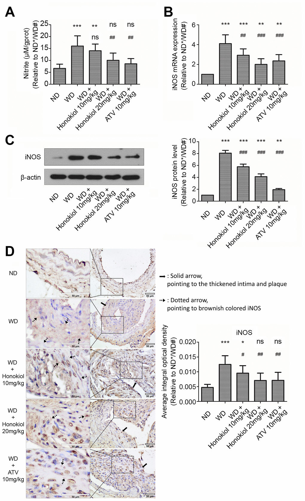
Figure 3.Effect of honokiol on NO production and iNOS expression in the carotid tissue of atherosclerotic mice. (A) The amount of nitric oxide in carotid tissue from the indicated experimental groups. (n = 6; * P < 0.05, ** P < 0.01, *** P < 0.001, vs. the ND group. # P < 0.05, ## P < 0.01, ### P < 0.001, vs. WD group; one-way ANOVA). (B) The mRNA expression of iNOS in carotid tissue obtained from the indicated groups was detected by real-time PCR. (n = 6; * P < 0.05, ** P < 0.01, *** P < 0.001, vs. the ND group. # P < 0.05, ## P < 0.01, ### P < 0.001, vs. the WD group; one-way ANOVA). (C) Western blotting was performed to evaluate iNOS protein expression in carotid tissue obtained from the indicated groups (upper panel). β-actin was served as a loading control. Quantification of the band density is shown in the right panel. (n = 6; * P < 0.05, ** P < 0.01, *** P < 0.001, vs. the ND group. # P < 0.05, ## P < 0.01, ### P < 0.001, vs. the WD group; one-way ANOVA). (D) Representative immunohistochemical staining images of iNOS in carotid tissue obtained from the indicated groups (hollow scale bar: 50μm for 400X; black solid arrows point to the thickened intima and plaque; black dotted arrows point to brownish colored α-SMA). The average integral optical density of iNOS in carotid tissue was quantitatively analyzed and shown in the right panel. (n = 6; * P < 0.05, ** P < 0.01, *** P < 0.001, vs. the ND group. # P < 0.05, ## P < 0.01, ### P < 0.001, vs. the WD group; one-way ANOVA). NO: Nitric oxide; iNOS: inducible nitric oxide synthase; ND: normal diet; WD: Western diet.