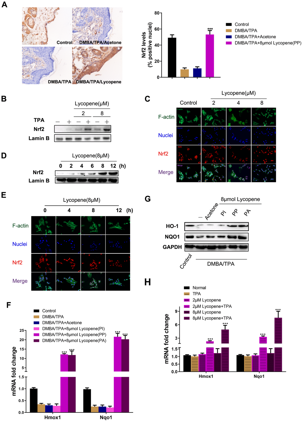
Figure 5.Lycopene activated the Nrf2 pathway in the presence of carcinogens in vivo and in vitro. (A) (Left panel) Representative images of Nrf2 immunohistochemistry staining in mouse epidermis in different groups (magnification 100×). (Right panel) Quantitative analysis of Nrf2 IHC results in left panel (n=9 per group). The data are presented as the mean ± SD. ***p < 0.001 (versus DMBA/TPA). (B) JB6 P+ cells were pretreated with increasing doses of lycopene for 12 hours and then exposed with or without TPA for additional 2 hours, and the nuclear levels of Nrf2 and LaminB1 were measured by Western blot. LaminB1 was used as the loading control. (C) JB6 P+ cells were pretreated with increasing doses of lycopene for 12 hours and then exposed with 20 ng/ml TPA for additional 2 hours. The immunofluorescence staining of Nrf2 was conducted as described in Materials and Methods (blue: nuclei, red: Nrf2, green: F-actin). (D) JB6 P+ cells were pretreated with 8 μM lycopene for various times and then exposed with TPA for additional 2 hours, and the nuclear levels of Nrf2 and LaminB1 were measured by Western blot. (E) The immunofluorescence staining of Nrf2. Treatment similar to (D). (F) Quantitative RT-PCR analysis of Nrf2 target genes in the mouse skin of the indicated groups (n=3). The data are presented as the mean ± SD. ***p < 0.001 (versus DMBA/TPA). (G) The levels of HO-1 and NQO1 in mouse skin were measured by Western blot. The results were representative of three independent experiments. (H) Treatment similar to (B), and mRNA levels of Hmox1 and Nqo1 were detected by real-time qPCR. GAPDH was used as the loading control. The data are presented as the mean ± SD. ***p < 0.001 (versus TPA alone).