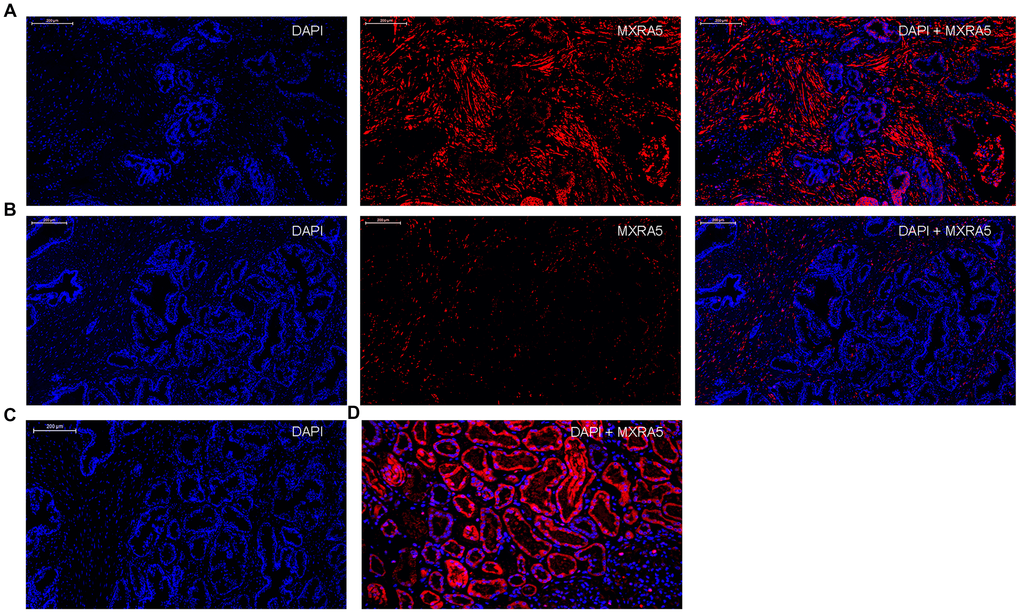
Figure 3.Immunofluorescence localization of MXRA5 in human prostate tissues. (A) Human BPH tissues. Left: DAPI (blue) indicates nuclear staining. Middle: Cy3-immunofluorescence (red) indicates the MXRA5 protein which was observed mainly in the fibromuscular stroma. Right: Merged image. (magnification ×200). (B) Human normal prostate. Left: DAPI (blue) indicates nuclear staining. Middle: Cy3-immunofluorescence (red) indicates MXRA5 protein. Right: Merged image (magnification ×200). (C) Negative controls omitting the primary antibody failed to stain. (D) Positive control using human renal cortex tissue showed a strong immune positivity for MXRA5 protein. DAPI (blue) indicates nuclear staining and Cy3-immunofluorescence (red) indicates MXRA5 protein staining (magnification ×200). Sections of all sample were used for immunofluorescence experiments and representative graphs were selected into figure.