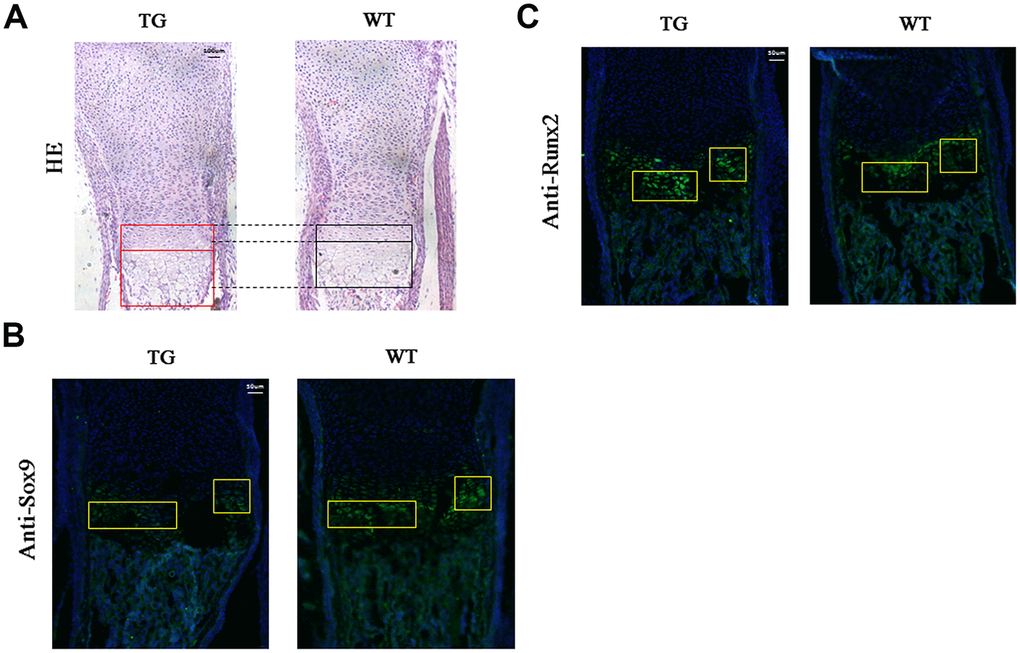
Figure 7.Histological and immunofluorescent analysis of Col10a1-TAp63γ transgenic mice. (A) The proliferative and hypertrophic zones of the limb cartilage in Col10a1-TAp63γ transgenic and WT mice were evaluated by hematoxylin and eosin (H&E) staining. Scale bar, 100 μm. (B) Sagittal sections of the distal humerus from both WT and transgenic mouse limbs at postnatal day 1 were subjected to immunofluorescent analysis using an anti-Sox9 antibody. Scale bar, 50 μm. (C) Sagittal sections of the distal humerus from both WT and transgenic mouse limbs at postnatal day 1 were subjected to immunofluorescent analysis using an anti-Runx2 antibody (label as Sox9 and some description of the findings). Scale bar, 50 μm.