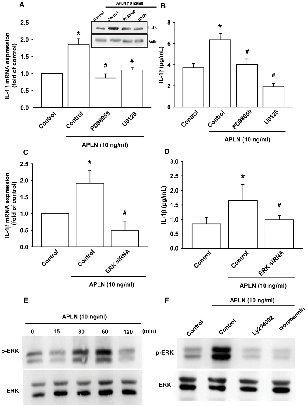
Figure 4.ERK phosphorylation is involved in APLN-induced IL-1β synthesis. (A) OASFs were pretreated with ERK inhibitors (PD98059, U0126; 10 μM) for 30 min, then incubated with APLN (10 ng/mL) for 24 h. IL-1β mRNA and protein levels were examined by RT-qPCR (n=4) and Western blot (n=3) assays, respectively. (B) OASFs were pretreated with ERK inhibitors (PD98059, U0126; 10 μM) for 30 min, then incubated with APLN (10 ng/mL) for 24 h. Excreted IL-1β protein levels were examined by ELISA (n=5). (C) OASFs were transfected with ERK siRNA (1 μg), then incubated with APLN (10 ng/mL) for 24 h. IL-1β mRNA levels were examined by ELISA assay (n=5). (D) OASFs were transfected with ERK siRNA (1 μg), then incubated with APLN (10 ng/mL) for 24 h. Excreted IL-1β protein levels were examined by ELISA assay (n=5). (E) OASFs were incubated with APLN (10 ng/mL) for the indicated time intervals, and the extent of ERK phosphorylation was examined by Western blot (n=3). (F) OASFs were pretreated with LY294002 and Wortmannin (10 μM) for 30 min, then incubated with APLN (10 ng/mL) for 24 h. The extent of ERK phosphorylation was examined by Western blot (n=3). * p<0.05 compared with control group; # p<0.05 compared with the APLN-treated group.