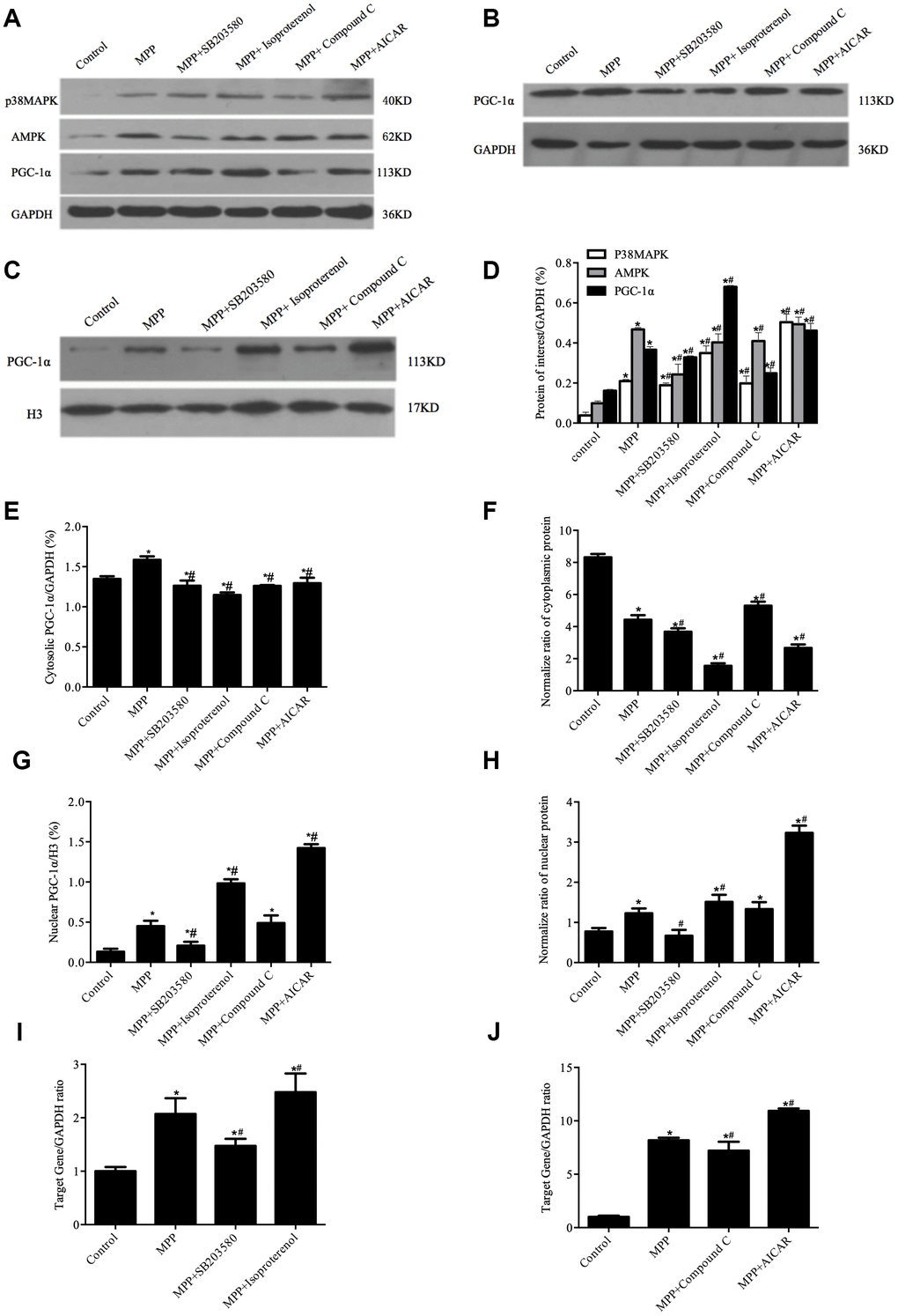
Figure 4.Redistribution of PGC-1α was regulated by p38MAPK and AMPK in MPP+-treated cell model. (A) Protein levels of p38MAPK, AMPK, and PGC-1α. (B, C) Cytosolic (B) and nuclear (C) protein levels of PGC-1α. (D) Semi-quantification of total protein levels of p38MAPK, AMPK, and PGC-1α relative to GAPDH; (E, G) Semi-quantification of cytosolic (E) and nuclear (G) protein levels of PGC-1α relative to GAPDH or H3; (F, H) Normalized cytosolic (F) and nuclear (H) proteins to the total proteins. (I, J) Transcriptional levels of p38MAPK (I) and AMPK(J) relative to GAPDH; n=6, per group. *P < 0.05, vs. Control group; # P < 0.05, vs.MPP+ group.