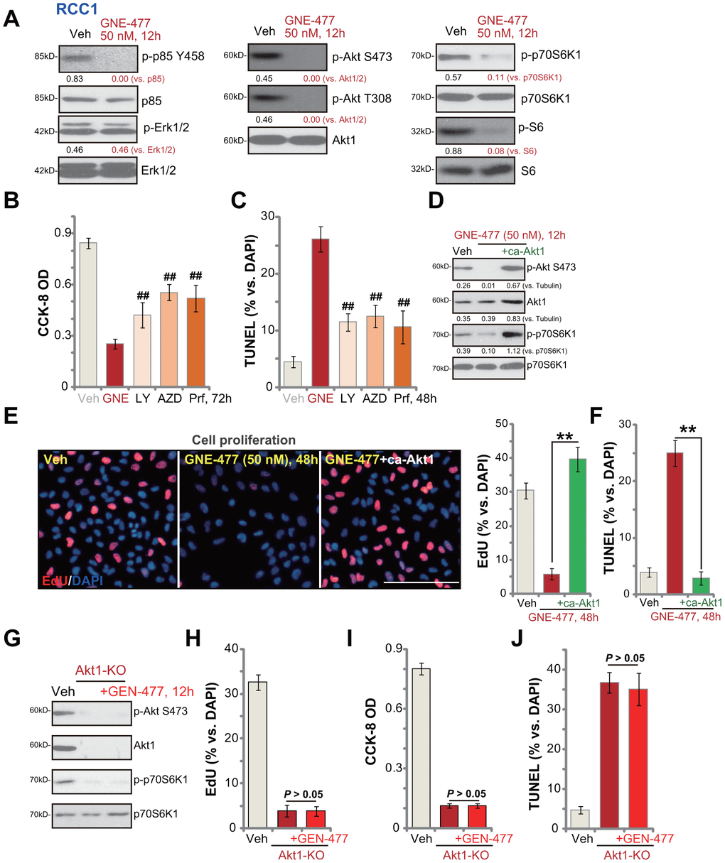
Figure 3.GNE-477 blocks PI3K-Akt-mTOR cascade activation in primary human RCC cells. RCC1 cells were treated with GNE-477 (50 nM) or the vehicle control (“Veh”, 0.1% DMSO), cells were further cultured for 12h, and expression of listed proteins tested by Western blotting (A); RCC1 cells were treated with GNE-477 (“GNE”, 50 nM), LY294002 (100 nM), AZD2014 (“AZD”, 100 nM), perifosine (“Prf”, 1 μM) or the vehicle control (“Veh”, 0.1% DMSO) for 48-72h, with cell viability and apoptosis tested by CCK-8 (B) and nuclear TUNEL staining (C) assays, respectively. The monoclonal stable RCC1 cells with or without the constitutively-active Akt1 (“+ca-Akt1”) construct were treated with GNE-477 (50 nM) or the vehicle control, cells were further cultured for applied time periods, expression of the listed proteins was tested (D); Cell proliferation and apoptosis were tested by EdU staining (E) and TUNEL assay (F), respectively. The monoclonal stable RCC1 cells with the CRISPR/Cas9 Akt1-KO construct (Akt1-KO cells) were treated with or without GNE-477 (50 nM), control cells with empty vector were treated with vehicle control (“Veh”), expression of listed proteins was shown (G); Cell viability, proliferation and apoptosis were tested by CCK-8 (H), EdU incorporation (I), and TUNEL staining (J) assays after 48h, respectively. Expression of listed proteins was quantified, normalized to the loading control (A, D). Bars stand for mean ± standard deviation (S.D.). For each assay, n=5. ##p < 0.01 vs. GNE-477 treatment (B, C). ** p < 0.01 (E, F). Experiments in this figure were repeated five times, and similar results obtained. Scale bar= 100 μm (E).