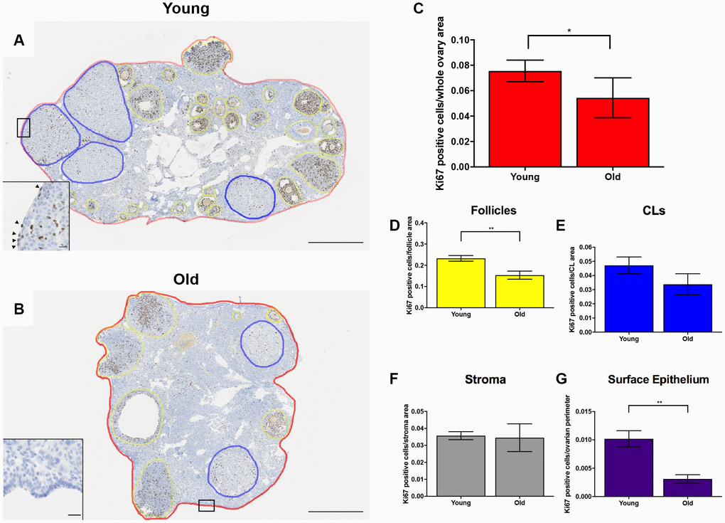
Figure 9.Cell proliferation is reduced post-ovulation in reproductively old mice. (A) Representative image of IHC labeling Ki67 in reproductively young ovaries. The whole ovarian area is outlined in red. Follicles are outlined in yellow. CLs are outlined in blue. (B) Representative image of IHC labeling Ki67 in reproductively old ovaries with the same ovarian sub-compartments outlined. Insets depict the boxed region of the OSE in each respective image. Scale bars are (A, B) 500 μm and (insets) 25 μm. Graphs showing the number of Ki67 positive cells per sub-compartment are within (C) whole ovaries, (D) follicles, (E) CLs, (F) the ovarian stroma, and (G) the OSE. T-tests were performed; asterisks indicate significant differences (C: P = 0.0294; D: P = 0.0091; G: P = 0.0023). Data are represented as mean ± SEM (C–G). N = 5 ovaries per age group.