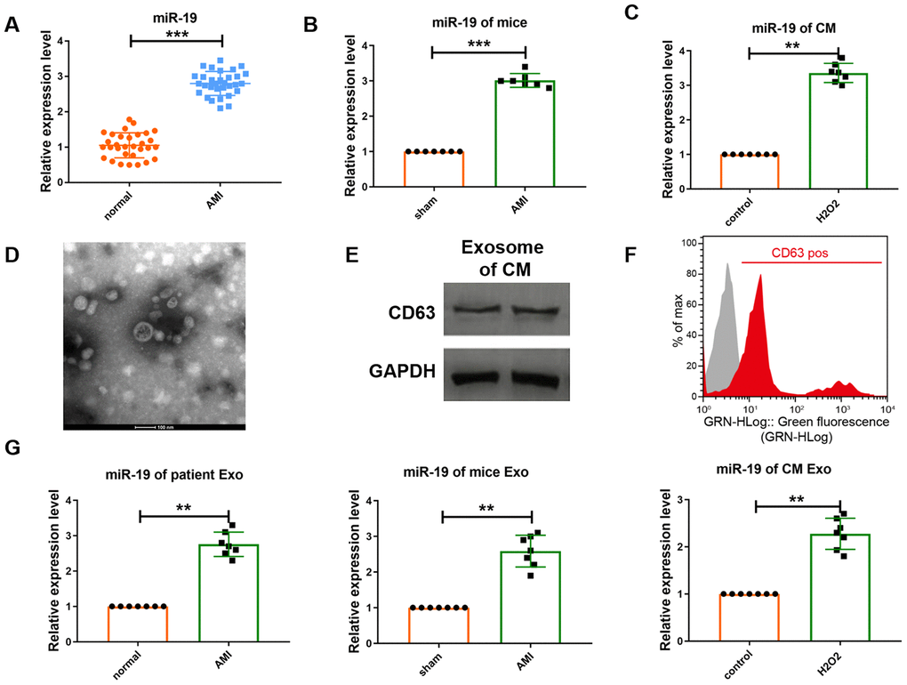
Figure 1.Expression level of miR-19a-3p is upregulated in response to MI or H2O2.. (A–C) qRT-PCR analysis of miR-19a-3p level in patients’ serum, mice serum and culture medium of CM. U6 was used as the control. (D) Electron microscopy image of CM–derived exosomes, showing a size of approximately 30 to 150 nm in diameter. Scale bar: 100 nm. (E) Western blot analysis of the protein level of CD63. (F) Flow cytometry analysis of CD63 of CM-derived exosomes. CM-derived exosomes were immunostained against CD63 (red curve) and compared with the appropriate isotype control (gray curve). (G) qRT-PCR analysis of miR-19a-3p level in exosomes derived from patients’ serum, mice serum and culture medium of CM. U6 was used as the control. **P < 0.01 and ***P < 0.001. All experiments were performed more than 3 biological repeats.