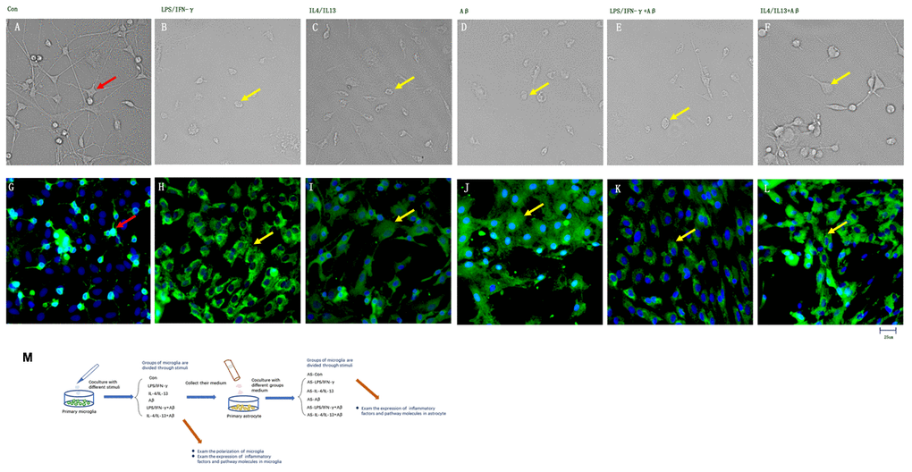
Figure 1.Inflammatory factors and Aβ activated primary microglia in vitro. (A–F) Microglial morphology was observed in light field images. (G–L) Microglia treated with different stimuli were stained with an anti-Iba1 antibody and observed on a fluorescence microscope. Green represents Iba1+ cells, while blue represents DAPI staining. (A, G) Control microglia. (B, H) Microglia treated with LPS/IFN-γ for 24 h. (C, I) Microglia treated with IL-4/IL-13 for 24 h. (D, J) Microglia treated with Aβ1-24 for 24 h. (E, K) Microglia treated with Aβ1-24 for 2 h, and then with LPS/IFN-γ for 24 h. (F, L) Microglia treated with Aβ1-24 for 2 h, and then with IL-4/IL-13 for 2 h. Scale bars, 25 μm. (M) Description of methods: Primary microglia were divided into six groups to be treated with different stimuli. Media from these microglia were then collected and co-cultured with primary astrocytes, and these astrocytes were divided into six groups according to the media with which they were treated. Microglial polarization was examined, and cytokine levels in both types of glia were measured.