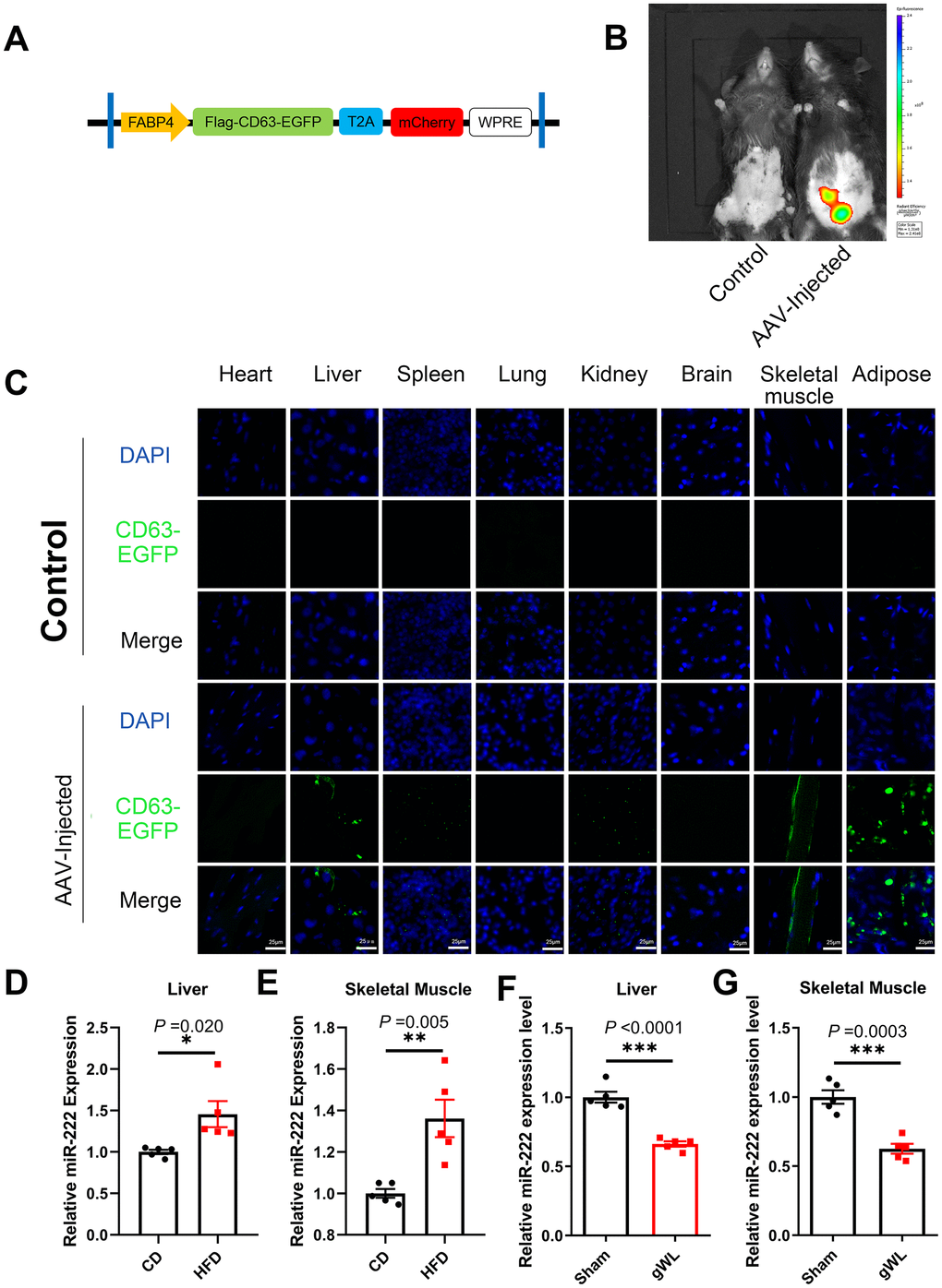
Figure 4.Liver and skeletal muscle are the major target tissues of gWAT-derived exosomal miR-222. (A) Diagrammatic representation of the AAV plasmid vector construct, HBAAV2/9-FABP4-3xflag-CD63-EGFP-T2A-mCherry. (B) Representative images show the in vivo imaging results in control (left) and AAV-injected (right) mice. The distribution of mCherry was detected. The expression of mCherry was observed in the gWAT of the AAV-injected mouse but was absent in the control. (C) Representative confocal microscopic images show the tissue distribution of the gWAT-derived exosomes that are marked by CD63-EGFP (green). The nuclei are stained using DAPI (blue). (D, E) QRT-PCR analysis results show the relative expression of miR-222 in the (D) liver and (E) skeletal muscle tissues from the CD-fed and HFD-fed mice (n=5 per group). (F, G) QRT-PCR analysis results show the relative expression of miR-222 in the (F) liver and (G) skeletal muscle tissues from gWAT-lipectomized (gWL) and sham-operated (Sham) HFD mice (n=5 per group). The data are presented as the means ± SE. * P < 0.05, ** P < 0.01, *** P < 0.001.