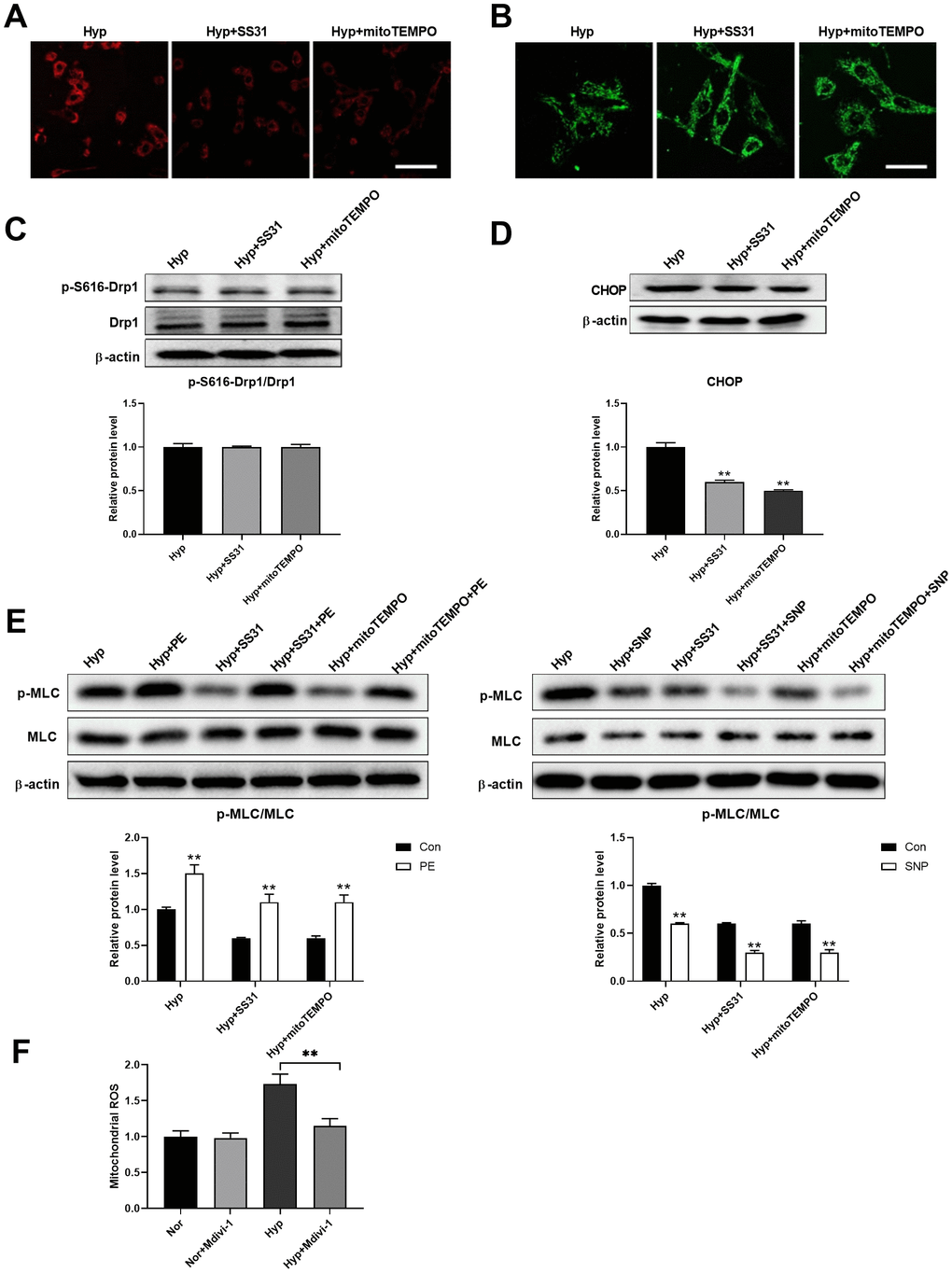
Figure 6.Mitochondrial ROS mediated the interaction between mitochondria and ER. (A) SS31 and mitoTEMPO scavenged mitochondrial ROS as detected by mitoSOX. Scale bar, 100 μm. (B) SS31 and mitoTEMPO inhibited hypoxia-induced mitochondrial fragmentation in PASMCs. Scale bar, 20 μm. (C) SS31 and mitoTEMPO showed little effects on Drp1 expression and Drp1 phosphorylation at serine 616 in PASMCs under hypoxia. Twenty micrograms of protein was loaded in each lane. (D) SS31 and mitoTEMPO decreased CHOP expression in PASMCs under hypoxia. Twenty micrograms of protein was loaded in each lane. (E) SS31 and mitoTEMPO improved PASMC function as evidenced by increased PE/SNP-induced MLC phosphorylation/dephosphorylation in PASMCs under hypoxia. Twenty micrograms of protein was loaded in each lane. (F) Inhibition of mitochondrial fragmentation using Mdivi-1 decreased mitochondrial ROS in PASMCs in hypoxia. *, p < 0.05, **, p < 0.01. n = 8.