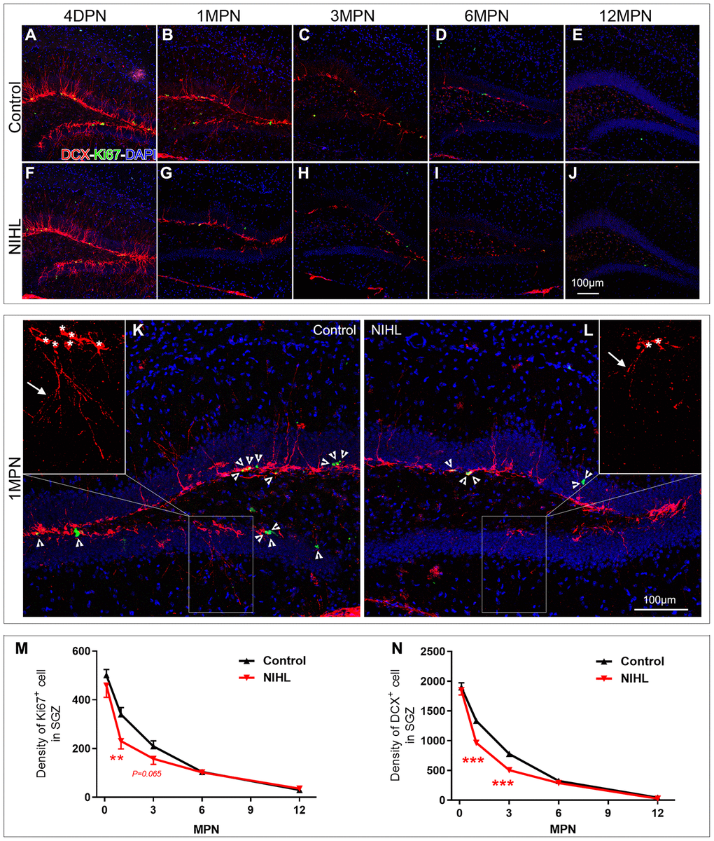
Figure 2.NIHL mice exhibited an accelerated age-related decline in hippocampal neurogenesis. (A–J) Representative images of Ki67+ (green) and DCX+ (red) cells in the hippocampal DG of control and NIHL mice at 4 DPN (A, F), 1 MPN (B, G), 3 MPN (C, H), 6 MPN (D, I) and 12 MPN (E, J). Note the accumulation of autofluorescent lipofuscin deposits (red), a sign of normal senescence [43], in the DG of control and NIHL mice at 6 MPN and 12 MPN. Scale bar: 100 μm. (K, L) Enlarged views of B (K, flipped horizontally) and G (L). Arrowheads indicate Ki67+ (green) cells. Asterisks and arrows in the magnified inserts indicate the soma and processes of DCX+ cells (red). Note the obvious reduction in branch complexity of DCX+ cells in NIHL mice. Scale bar: 100 μm. (M, N) Quantitative analyses of Ki67+ cells (M) and DCX+ cells (N) in the DG of mice at different time points. **P<0.01, ***P<0.001 (two-way ANOVA, post hoc Tukey’s test, vs. age-matched control group).
Figure 2 — Accelerated age-related decline in hippocampal neurogenesis in mice with noise-induced hearing loss is associated with hippocampal microglial degeneration | Aging