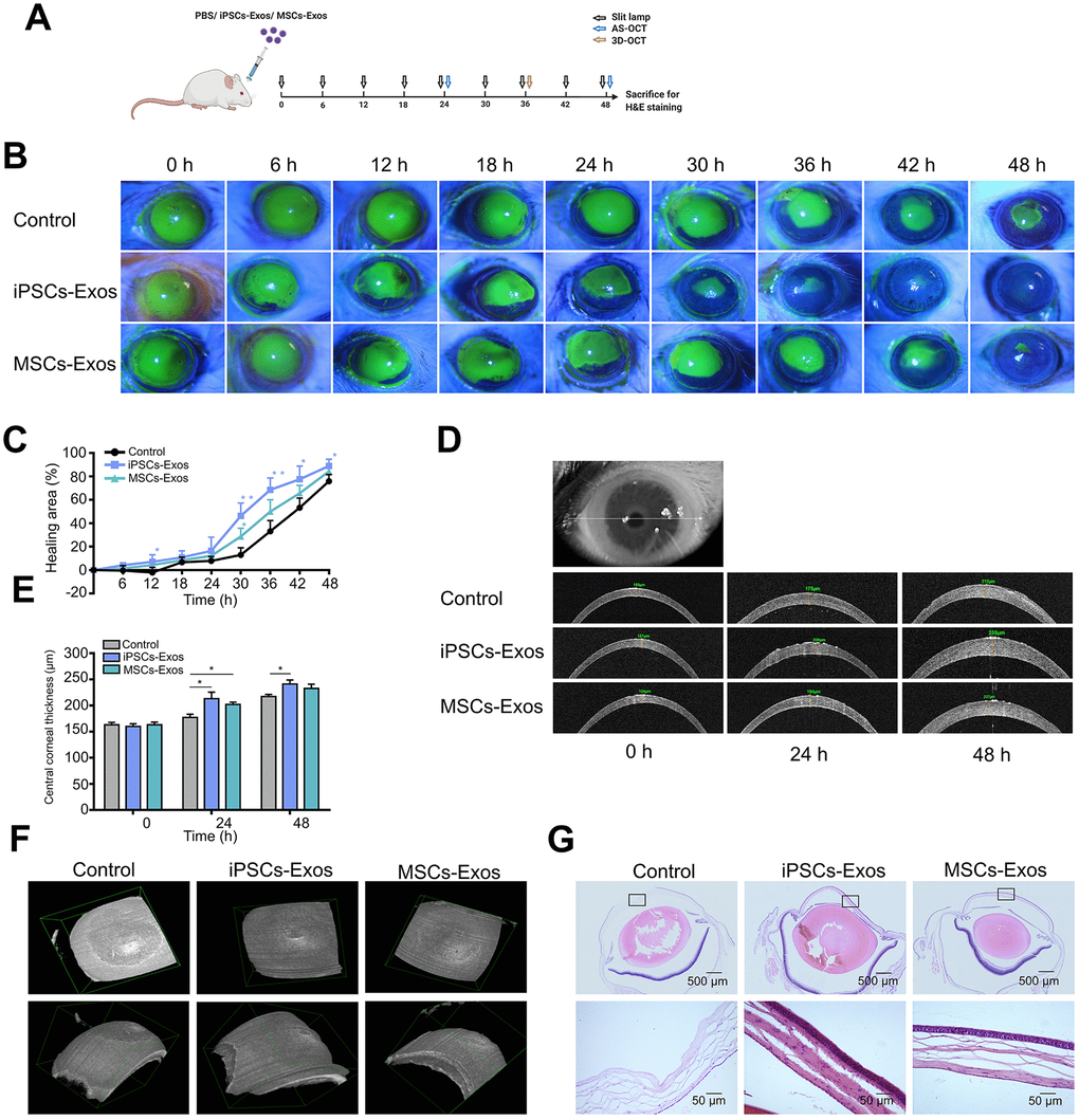
Figure 5.Effect of iPSCs/MSCs-Exos on corneal epithelial defect healing in vivo. (A) Schematic diagram of the experimental procedure. After corneal epithelial defect and treated with iPSCs/MSCs-Exos every 6 h, different procedures were performed at relative time point. (B) The corneal epithelial defect area was monitored every 6 hours with fluorescein staining and slit lamp. (C) The corneal epithelial defect healing rates are shown in line graphs. The cornea healing area in iPSCs-Exos group increased obviously compared with that in the vehicle group and was statistically significant at 12, 30, 36, 42, and 48 h compared with control group. (D) Central corneal thickness was measured by AS-OCT at both 24 and 48 h. (E) Central corneal thickness is shown by bar graph. Central corneal thickness in the iPSCs-Exos group was statistically different from that of the control. (F) A 3D-pattern was generated by AS-OCT for rat corneas at 36 h, enabling a more stereoscopic view of the corneal epithelial defects. (G) Rat eyeballs were harvested 48 h after corneal defect and were stained with hematoxylin and eosin. Data are representative of one of three independent experiments performed. Each experiment consisted of 4 animals/group. The values are shown as the mean ± SEM. * P<0.05, ** P<0.01.