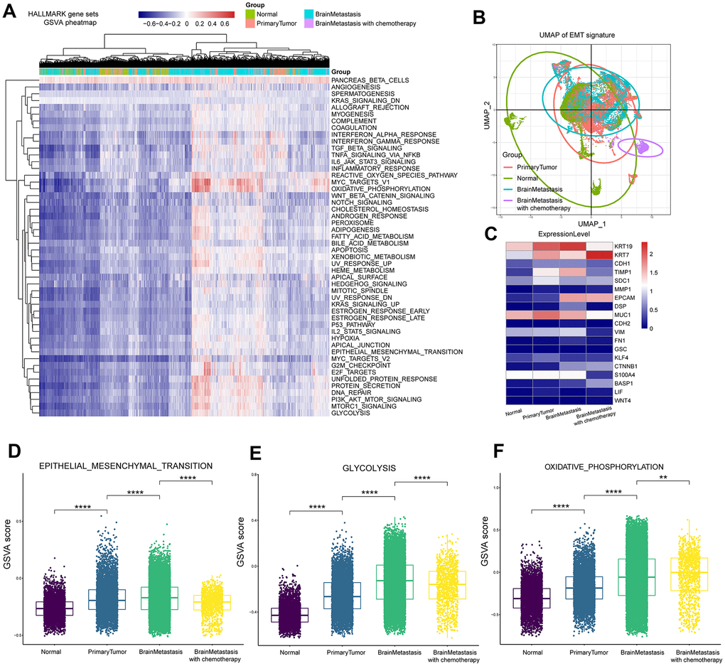
Figure 4.Gene set variant analysis of LUAD patient samples from primary tumors and brain metastases. (A) Heatmap of 50 cancer hallmark gene sets in primary LUAD and brain metastasis samples. The color index from navy blue to red indicates low to high expression of the gene sets. (B) The UMAP graph shows the diversity of EMT gene expression in the primary LUAD and brain metastasis samples at different stages of cancer progression. (C) Heatmap shows the mean expression of EMT-associated genes in the primary LUAD and brain metastasis samples at different stages of cancer progression. (D) The box plot shows the expression of the EMT pathway genes in the primary LUAD, brain metastasis (with or without chemotherapy) samples. (E) The box plot shows the expression of the glycolysis pathway genes in the primary LUAD and brain metastasis (with or without chemotherapy) samples. (F) The box plot shows the expression of the oxidative phosphorylation pathway genes in the primary LUAD and brain metastasis (with or without chemotherapy) samples