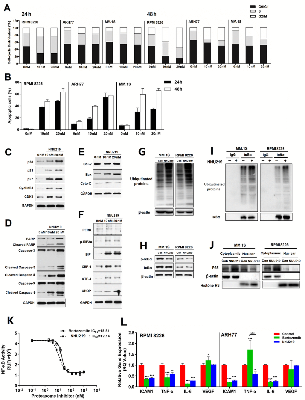
Figure 3.NNU219 induces apoptotic signal transduction and modulates the NF-κB signaling pathway in human MM cell lines. (A) The MM cell lines RPMI 8226, ARH77 and MM.1S were incubated with DMSO or NNU219 (10 and 20 nM) for 24 or 48 h, respectively. Cells were fixed with ethanol and stained with propidium iodide, then DNA content was determined by flow cytometry (p < 0.05 for all cell lines). (B) The MM cell lines RPMI 8226, ARH77 and MM.1S were incubated with DMSO or NNU219 (10 and 20 nM) for 24 or 48 h and induction of apoptosis was determined after annexin V-FITC/propidium iodide staining by flow cytometry. Data were presented as mean ± SD of three independent experiments (p < 0.001 for all cell lines). (C–F) RPMI 8226 cells were incubated with NNU219 (10 and 20 nM) for 24 h. After the incubation, cells were lysed and directly subjected to SDS-PAGE, transferred to membranes and blotted with indicated antibodies. GAPDH immunoblotting was included for protein loading control. Blots in the figures were representatives of three independent experiments. (G) MM1.S and RPMI 8226 cell lines were treated with 10 nM of NNU219 for 24 h. The stimulated cells were lysed and 20 μg of protein was processed for ubiquitin immunoblotting. (H) MM1.S and RPMI 8226 cells were treated with DMSO, 10 nM of NNU219 for 24 h and immunoblotted using anti-IκBα and anti-phospho-IκBα antibodies. (I) MM1.S and RPMI 8226 cell lines were treated with 10 nM of NNU219 for 24 h. Correlative proteins were immunoprecipitated from 1 mg of MM cell lysate using IκBα or IgG antibody (rabbit) and coupled to protein A/G agarose beads. The beads were washed by IP buffer and processed by immunoblotting for ubiquitin, p65 or IκBα. (J) MM1.S and RPMI 8226 cell line was treated with 10 nM of NNU219 for 24 h. Nuclear and cytoplasmic protein fractions were separated from the total lysate and analyzed for p65, β-actin and histone antibodies by western blot analysis. Blots in the figure were representative of three independent experiments. (K) The transfected NF-κB/Luciferase 293T cells were incubated with increasing concentrations of NNU219 or bortezomib for 6 h. Cells were then stimulated with 10 ng/ml of TNF-α for another 18 h. The activity of expressed luciferase was determined by using the Dual-Luciferase Reporter Assay System. (L) RPMI 8226 or ARH77 cell lines were treated with DMSO, IC50 of NNU219 or bortezomib for 12 h and harvested. Total RNA was isolated and subjected to qRT-PCR. The gene expression level of ICAM1, TNF-α, IL6 and VEGF was normalized to GAPDH using the 2−ΔΔCT method. In above experiments, the control group was incubated with the same concentration of DMSO in normal culture medium. Values were expressed as mean ± SD of triplicate samples of three independent experiments (*, p < 0.05; **, p < 0.01; ***, p < 0.001). IL, interleukin; TNF, tumor necrosis factor; ICAM, intercellular adhesion molecule; VEGF, vascular endothelial growth factor; p-IκBα, phosphorylated inhibitor of NF-κB; Con, control; MM, multiple myeloma.