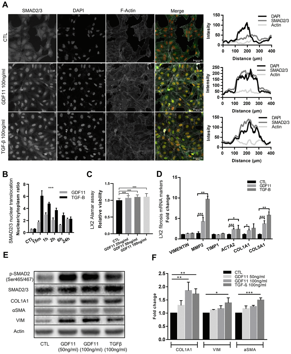
Figure 6.Stellate cells are activated and produce ECM components after GDF11 exposure. (A) Activation of the LX2 stellate cell line by GDF11 (100 ng/ml) or TGF-β (100 ng/ml, positive control) (scale= 100μm). (B) The nuclear translocation ratio of SMAD2/3 complexes after GDF11 and TGF-β treatment (n=3 per group). (C) Cell viability was assessed in LX2 cells, treated with different doses of GDF11 (25, 50, 100 ng/ml) for 48 hours, by using alamarBlue™ Cell Viability Reagent. (D) Relative mRNA expression of liver fibrosis/stellate cells activation markers in LX2 cells after exposure to GDF11 (100 ng/ml) or TGF-β (100 ng/ml). (E) Protein expression levels of liver fibrosis/stellate cells activation markers in LX2 cells after GDF11 or TGF-β exposure. Protein expression of selected effectors (pSMAD2, COL1A1, αSMA, vimentin (VIM), actin, GAPDH) were quantitatively assessed by immunoblotting in LX2 cells exposed or not for 48 h to GDF11 (50 or 100 ng/ml) or stimulated for 48h with TGF-β (100 ng/ml). Images are representative of three independent experiments. (F) Data quantification represents the means ± SD. * p<0.05; ** p<0.01; *** p<0.001 (Mann-Whitney U-test).