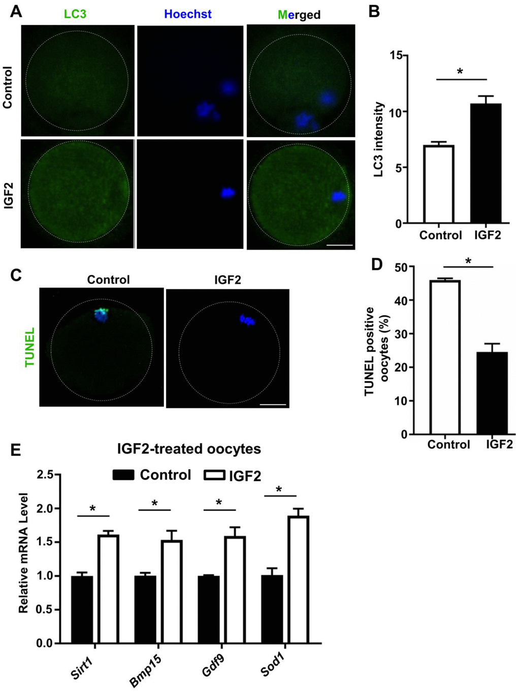
Figure 6.IGF2 reduces the apoptosis and promotes the level of autophagy in aged mouse oocytes. (A) LC3 staining showing the extent of autophagy occurring in control and IGF2-treated oocytes. (B) Quantification of LC3 intensity in control (n = 34) and IGF2-treated oocytes (n = 25). A Student’s t-test (two-tailed). *p < 0.05. Error bars indicate the SEM. (C) TUNEL assay of control and IGF2-treated oocytes from aged mice. A green fluorescence signal indicates TUNEL-positive oocytes. Apoptotic signals were observed after 16 h of in vitro culture. DNA was counterstained with DAPI. Scale bar = 30 μm. (D) The percentage of apoptosis-positive oocytes in control (n = 61) and IGF2-treated oocytes group (n = 44). A Student’s t-test (two-tailed). *p < 0.05. Error bars indicate the SEM. (E) qPCR results showing mRNA levels of Sirt1, Bmp15, Gdf9, and Sod1 in MII-stage oocytes after in vitro maturation with or without IGF2-treatment. *p < 0.05. A Student’s t-test (two-tailed). Error bars indicate the SEM.