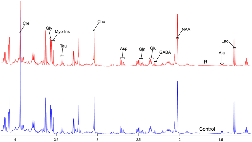
Figure 2.The normalized 1H NMR spectra of extracts in the parietal cortex after MIRI.
Figure 2 — Neurochemical alterations of different cerebral regions in rats with myocardial ischemia-reperfusion injury based on proton nuclear magnetic spectroscopy analysis | Aging