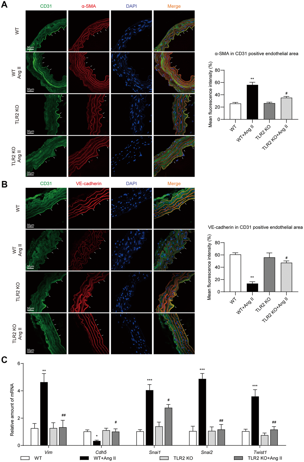
Figure 2.TLR2 KO mice are protected against Ang II-induced EndMT in aortas. (A) Representative immunofluorescence staining images showing CD31 (green) and α-SMA (red) in mouse aortic tissues. Arrows indicate positive staining. Tissues were counterstained with DAPI (blue) [scale bar = 30 μm]. Quantification of α-SMA fluorescence in CD31-positive endothelial area is shown on right. (B) Representative immunofluorescence staining images showing CD31 (green) and VE-cadherin (red) in mouse aortic tissues. Arrows indicate positive staining. Tissues were counterstained with DAPI (blue) [scale bar = 30 μm]. Quantification of VE-cadherin in CD31-positive endothelial area is shown on right. (C) mRNA levels of EndMT-associated genes in aortas [Data normalized to β-actin]. [n = 6-8; Data shown as Mean ± SEM; *p<0.05, **p<0.01, and ***p<0.001 compared to WT; #p<0.05 and ##p<0.01 compared to WT-Ang II].