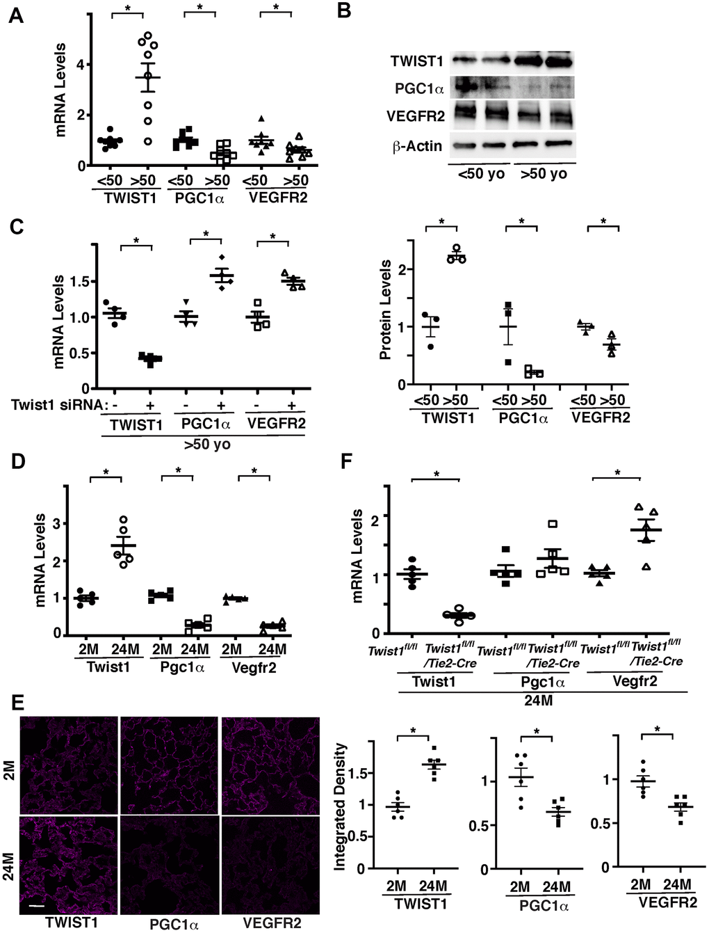
Figure 1.Twist1 mediates age-dependent decline in PGC1α and VEGFR2 expression in ECs. (A) Graph showing the mRNA levels of Twist1, PGC1α, and VEGFR2 in ECs isolated from young (<50 years old) vs. old (>50 years old) human adipose tissues (n=8, mean ± s.e.m., *, p<0.05). (B) Representative immunoblots showing Twist1, PGC1α, VEGFR2, and β-actin protein levels in young vs. aged human adipose ECs (top). Graph showing Twist1, PGC1α, and VEGFR2 protein levels normalized by β-actin protein levels in young vs. aged human adipose ECs (bottom, n=3, mean ± s.e.m., *, p<0.05). (C) Graph showing the mRNA levels of Twist1, PGC1α, and VEGFR2 in aged human adipose ECs treated with Twist1 siRNA or control siRNA with irrelevant sequences (n=4, mean ± s.e.m., *, p<0.05). (D) Graph showing the mRNA levels of Twist1, Pgc1α, and Vegfr2 in ECs isolated from 2M vs. 24M old mouse lungs (n=5, mean ± s.e.m., *, p<0.05). (E) IF micrographs showing Twist1, PGC1α, and VEGFR2 expression in 2M vs. 24M old mouse lungs. Scale bar, 50 μm. Graphs showing integrated density of Twist1, PGC1α, and VEGFR2 in the lung tissues (n=6, mean ± s.e.m., *, p<0.05). (F) Graph showing the mRNA levels of Twist1, Pgc1α, and Vegfr2 in ECs isolated from 24M old Twist1fl/fl and Twist1fl/fl/Tie2-cre mouse lungs (n=5, mean ± s.e.m., *, p<0.05).