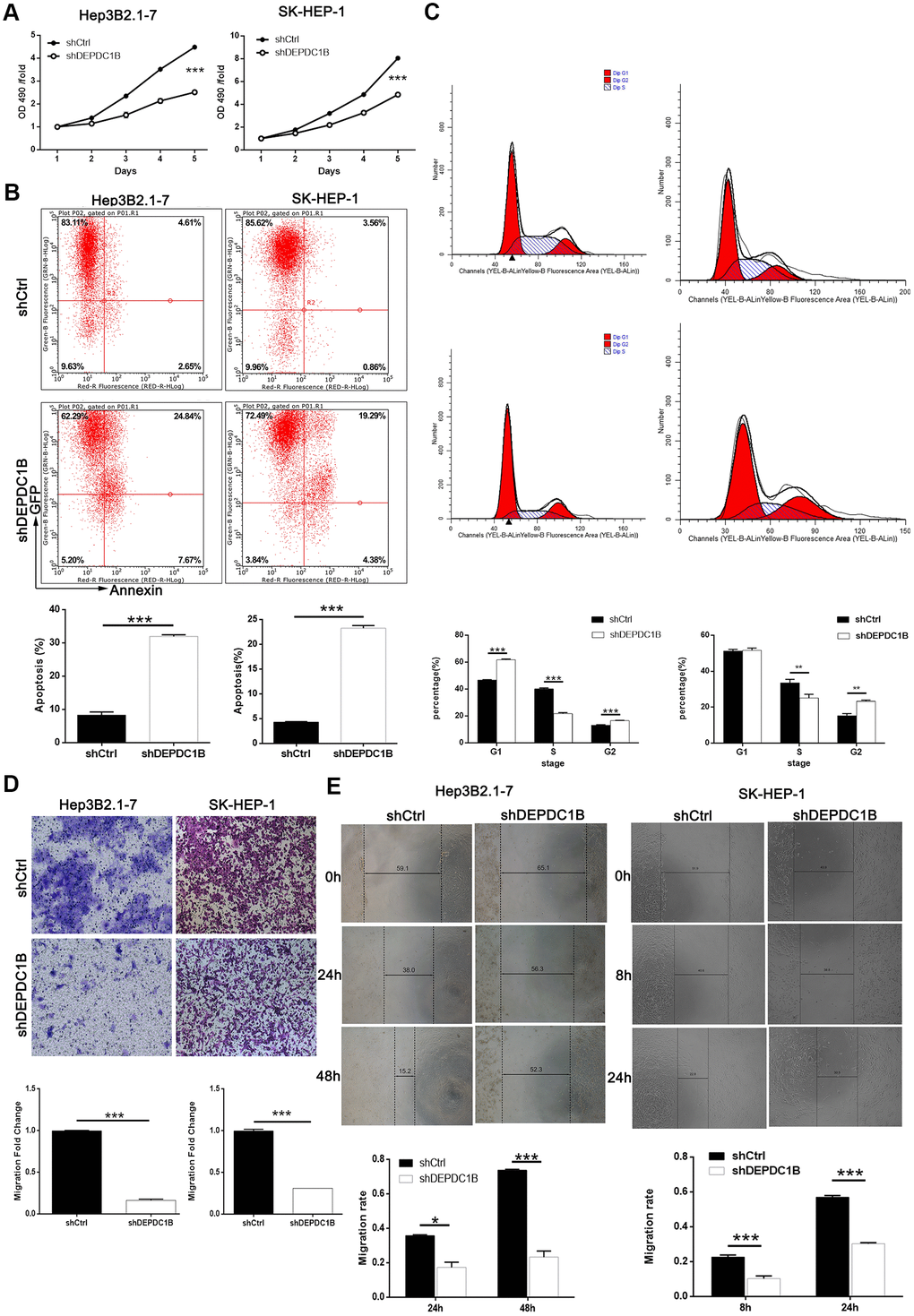
Figure 3.DEPDC1B knockdown inhibited HCC cell progression in vivo. (A) The results of MTT assay show that, after the infection of lentivirus: compared with shCtrl group, the cells in shDEPDC1B group exhibited slower proliferation rate (P<0.001). (B) The results of flow cytometry demonstrate that, after the infection of lentivirus: compared with shCtrl group, apoptosis percentage was increased in shDEPDC1B group (P<0.001). (C) The results of flow cytometry show that, compared with shCtrl group, in shDEPDC1B group, the percentage of SK-HEP-1 cells in G2 phase increased (P<0.01), and the percentage of Hep3B2.1-7cells in G2 phase increased significantly in shDEPDC1B group (P<0.001). Transwell assay showed that, after the infection of lentivirus: compared with shCtrl group, the migration ability of cells in shDEPDC1B group was inhibited (P<0.001) (D). (E) The results of wound-healing assay showed that, in HEP3B2.1-7 and SK-HEP-1cells, compared with shCtrl group, the migration rate of cells in shDEPDC1B group was decreased by 68% (P<0.001) and 47% (P<0.001). **: P <0.01. ***: P <0.001.