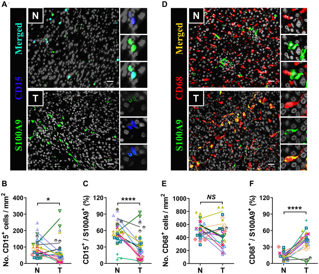
Figure 6.Myeloid cells are the major source of S100A9. Multiple immunofluorescence staining shows DAPI (gray), S100A9 (green), CD15 (blue, A), and CD68 (red, D) expression and coexpression (double-positive cells) in the N and T regions. Quantification of CD15+ (B) and CD68+ (E) cell densities in the T and N regions (n = 12). (C) The percentages of S100A9+CD15+ cells among the total S100A9+ cells in the N and T regions. (F) The percentages of S100A9+CD68+ cells among the total S100A9+ cells in the N and T regions. (n = 12). Scale bar = 25 μm. N, nontumor, T, tumor. *P < 0.05, ****P < 0.0001, NS, no significance.