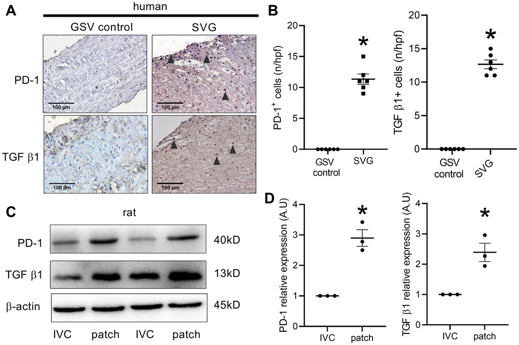
Figure 1.PD-1 expression in human and rat venous neointimas. (A) Immunohistochemistry images showing PD-1 and TGF β1 expression in the native human great saphenous vein (GSV control) and the neointima of spiral great saphenous vein graft (SVG); black arrowhead showing positive cells; scale bar, 100 μm; n = 3. (B) Bar graphs showing PD-1- (*p < 0.0001, t-test) and TGF β1-positive cells (*p < 0.0001, t-test) per high-power field in the human GSV and SVG neointima, n = 6. (C) Western blot showing the expression of PD-1, TGF β1, and β-actin in the rat IVC and the patch after patch venoplasty at day 14; n = 3. (D) Bar graph showing PD-1 (*p = 0.0022, t-test) and TGF β1 (*p = 0.0128, t-test) density; n = 3.