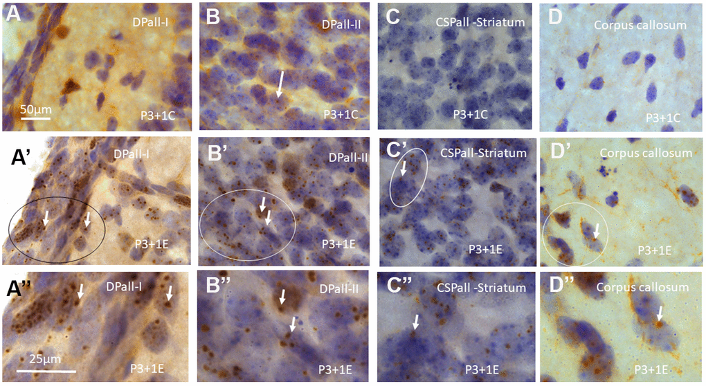
Figure 1.γH2AX immunostaining counterstained with hematoxylin shows very few γH2AX foci in different brain regions of postnatal day 4 (P4) mice without irradiation including dorsal pallium(DPall) /isocortex layer I (DPall-I) and pia mater (A), DPall –II (B, arrow), central subpallium/classic basal ganglia (CSPall) striatum (C) and corpus callosum (D). However, acute irradiation with 5Gy at P3 induced very significant γH2AX expression in the entire brain 1 day after radiation exposure or P4 mice. γH2AX foci could be observed in almost all brain regions at 1 day after irradiation at P3, including DPall-I (A’, A” is magnified from the ellipse in A’), DPall-II to DPall-VI of the grey mater (B’, B”, DPall-II, B” is magnified from the ellipse in B’), CSPall striatum (C’, C” is magnified from the ellipse in C’) and corpus callosum (D’, D” is magnified from the ellipse in D’) at 1 day after irradiation at P3. Scan bar=50μm in (A) applies to (B–D) (A’–D’) Scan bar=25μm in (A”) applies to (B” –D”).