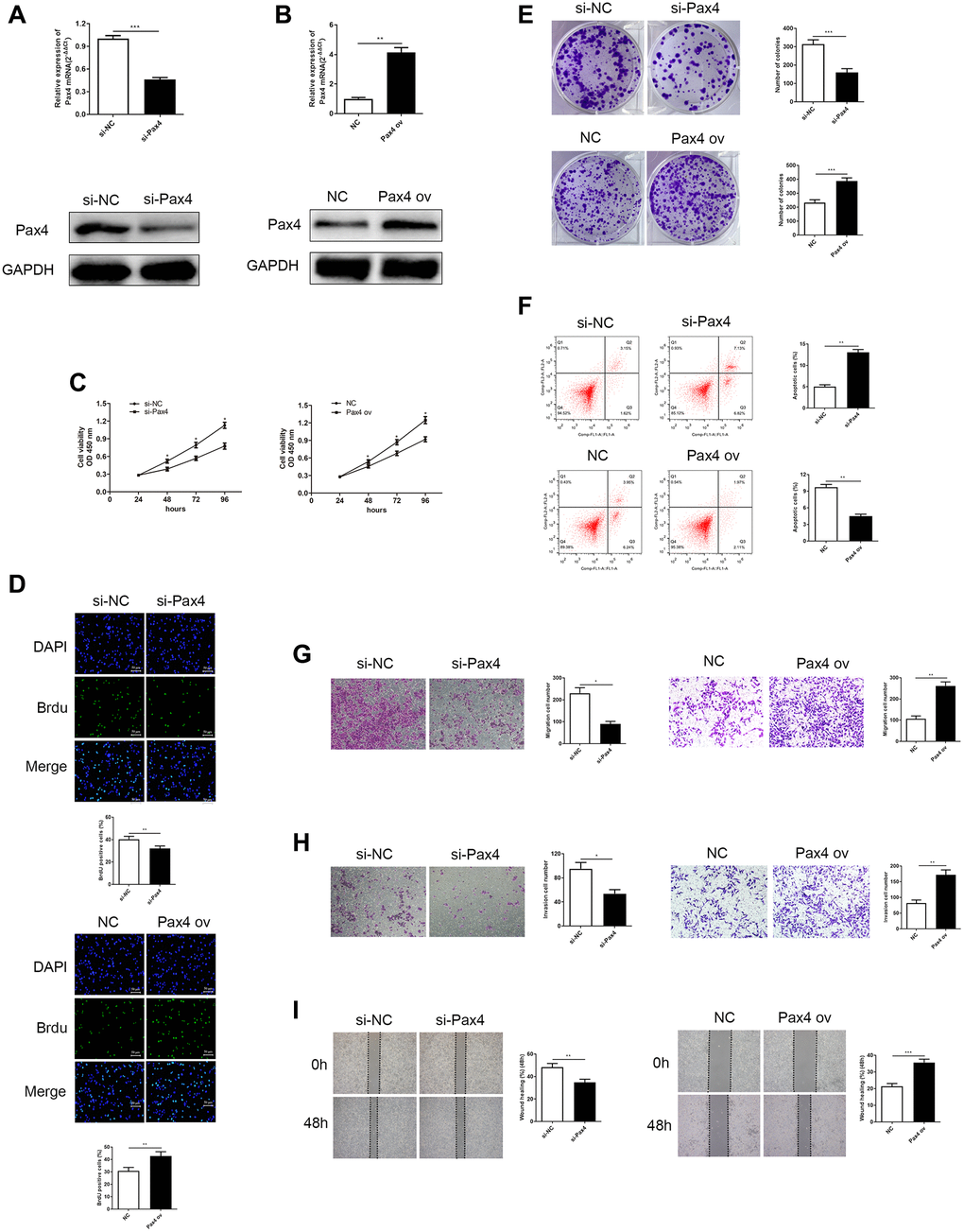
Figure 2.Knockdown of PAX4 in AGS cells inhibited GC cell proliferation, migration and invasion, while overexpression of PAX4 in the HGC-27 cells accelerates GC proliferation, migration and invasion. (A) PAX4 mRNA and protein levels were decreased after si-PAX4 transfection in AGS cells compared to the control group (P = 0.0004). (B) PAX4 mRNA and protein levels were increased after PAX4 ov transfection in HGC-27 cells compared to the NC group (P = 0.0058). (C) Lower PAX4 expression weaken GC cell viability and higher PAX4 expression was beneficial for GC cell viability assessed by the CCK-8 assay. (D) Lower PAX4 expression weaken GC cell proliferation (P = 0.0016) and higher PAX4 expression was beneficial for GC cell proliferation (P = 0.0091) assessed by the Brdu assay. (E) Colony formation assay determined numbers of colony formation after si-PAX4 (P = 0.0005) or PAX4 ov (P = 0.0005) transfected (P = 0.0005). (F) Flow cytometry assay demonstrated that GC cell apoptosis capacity was strengthened in the absence of PAX4 (P = 0.0014) and was attenuated in PAX4 ov group (P = 0.0011). (G–H) Transwell assay demonstrated that decreased PAX4 result in inhibition of AGS cell migration (P = 0.012) and invasion (P = 0.013), increased PAX4 enhanced GC cell migration (P = 0.0014) and invasion (P = 0.0041). (I) Wound healing assay indicated the impaired GC cell migration when reduced PAX4 (P = 0.0076) and fortified GC cell migration when PAX4 was overexpressed (P = 0.0005).