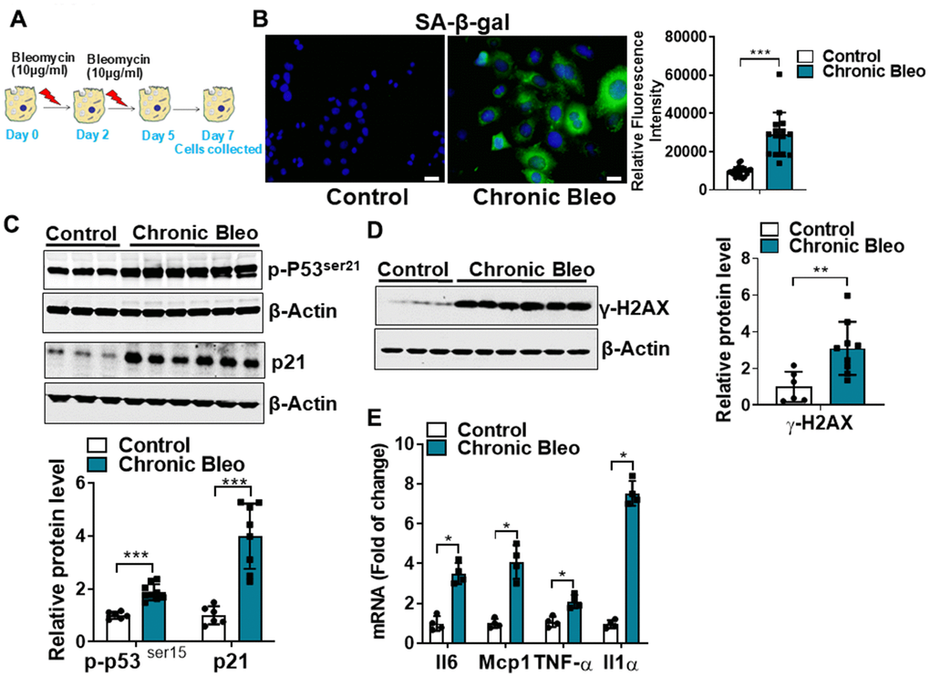
Figure 1.Cellular senescence markers are increased in MLE12 cells after our 7-day injury protocol. (A) Schematic showing the experimental design: On day 0, MLE-12 cells were treated with 10 μg/ml bleomycin for 24h. Day 2, bleomycin was removed, and the culture medium was refreshed. Day 4, cells were treated again with 10 μg/ml bleomycin. Day 5, bleomycin was subsequently removed, and cells were collected at day 7. (B) SA-β-gal activity (green fluorescence cytoplasmic staining) in control and chronically injured MLE12 cells. Number of SA-β-gal positive cells per 100 cells counted (right). (C) Western blot (WB) for p-p53Ser21 and p21 in chronically injured lung epithelial cells (with β-actin loading control). Densitometry is shown on the right. (D) WB for γ-H2AX in control and bleomycin injured cells (with β-actin loading control). Densitometry is shown on the right. (E) Transcript levels for Il6, Mcp1, Tnf-α, and Il1α in controls vs. chronically injured lung epithelial cells. Statistical significance was assessed by unpaired Student’s t-test for two groups. * p< 0.05, **P < 0.01, ***P < 0.001 versus control group, n=6.